


Department of Dermatology, Aarhus University Hospital, DK-8000 Aarhus, Denmark. E-mail: temkris@post.au.dk
Accepted May 29, 2019; E-published May 29, 2019
Psoriasis is a chronic inflammatory skin disease characterized by scaly well-demarcated plaques and a high comorbidity burden. Disease management is complex and should include a patient-centred view (1). In Denmark, Dead Sea climatotherapy (DSC) has been used successfully for many years as a treatment approach to psoriasis, with excellent clearance rates (2). It is generally believed that the cutaneous symptoms of psoriasis often relapse in previously affected sites after cessation of treatment, although this has not been documented or quantified. Relapse indicates the presence of a “molecular scar” in clinically healed skin (3). The aim of this study was to determine whether recurrence of psoriasis after complete clearance occurs in a non-random fashion. This was determined by automated imaging in patients achieving complete clearance after treatment with DSC.
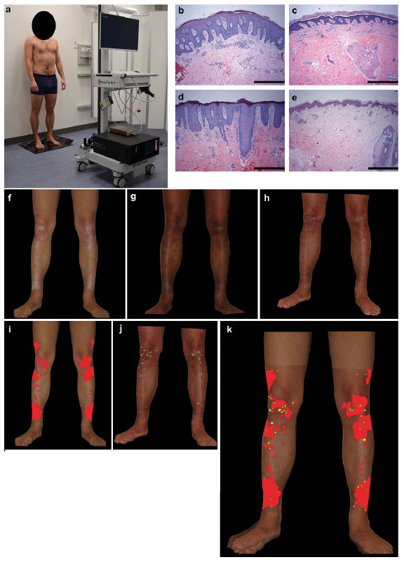
Six patients with moderate to severe plaque-type psoriasis were included. All patients signed an individual written informed consent to participate, and study approval was obtained from The Central Denmark Region Committees on Health Research Ethics. Procedures were conducted in accordance with ethical standards on human experimentation and the Declaration of Helsinki 1975, as revised in 1983. Clinical improvement was evaluated by the reduction in the Psoriasis Area and Severity Index (PASI) and the 5-point Investigator’s Global Assessment (IGA). Automated Total Body Mapping (ATBM) images were acquired using the FotoFinder Body-studio ATBM (FotoFinder Systems GmbH, Bad Birnbach, Germany) (Fig. 1a). A digital reflex camera (Canon EOS 700D; Canon, Lake Success, NY, USA; with Canon EF-S 18-55mm f/3.5-5.6 IS STM lens; Canon, Lake Success, NY, USA) was attached to a motor-powered vertical rail. Camera movement was powered by an automatic camera positioning system. Patients were encouraged to assume different poses for automatic image acquisition. Four pictures from 4 different views (frontal-, left sagittal-, right sagittal- and dorsal plane) were acquired, creating a total of 16 pictures per patient. Digital images were transferred to Adobe Photoshop® CC 2018 (Adobe, Park Avenue, San Jose, CA, USA). Furthermore, a 4-mm punch biopsy specimen was acquired from the same target lesion of a psoriatic plaque at baseline (Fig. 1b), visit 1 (Fig. 1c) and visit X (Fig. 1d), and from non-lesional psoriatic skin at baseline (Fig. 1e). Biopsies were stored and stained with haematoxylin and eosin (HE), as described previously (4). Fig. 1f–h shows representative pictures from the same patient obtained at baseline, visit 1 and visit X, respectively. The lesional area was marked manually at baseline (Fig. 1i), visit 1 (Fig. 1j) and the number of pixels was counted. Pictures from baseline and visit X were then overlaid, and the overlapping area was calculated (Fig. 1k). Subsequently, for each picture a ratio was calculated, based on both the overlapping lesional area between baseline and visit X and the lesional area compared with body surface area (BSA). The feet, hands, genital area, intertriginous areas, head and non-overlapping areas were not included in the BSA.
Fig. 1. (a) Automated Total Body Mapping (ATBM) setup. Representative haematoxylin and eosin (HE)-stains originating from the same target lesion of the same patient: (b) at baseline, (c) immediately after, and (d) at first visible signs of psoriasis. (e) HE-staining of non-lesional skin. Scale bars: 250 µm (original magnification x 100). Reversal of psoriatic epidermal features after Dead Sea climatotherapy. (f) Representative clinical photographs from the same patient at baseline, (g) immediately after returning from Dead Sea climatotherapy, and (h) at first visible signs of psoriasis. (i and j) Plaque area painted manually at baseline and visit X, respectively. (k) Overlapping area in green. Written permission is provided by the patient.
The patients comprised 5 males and 1 female. At baseline and visit X mean PASI was 13.9 (standard deviation (SD) 5.5, range 8.08–19.6) and 4.53 (SD 2, range 1.34–7.71), respectively. All patients enrolled in this part of the study should have an IGA and PASI of 0 at visit 1. The results showed that 60.2% (SD 23.6%, range 34.7–94.3%) of the new plaque area reappeared inside the location of former plaques. At baseline and at visit X, 14.1% (SD 17, range 1.56–46.45%) and 8.2% (SD 16.4%, range 0.07–41.58%), respectively, of the BSA was covered by visible plaques. Histopathology examination showed typical psoriatic features at baseline (Fig. 1b). Immediately after treatment with DSC (Fig. 1c), a normal phenotype was seen, similar to non-lesional skin (Fig. 1e). Histopathological examination at visit X confirmed the reappearance of psoriasis (Fig. 1d).
This is the first study to investigate the plaque-site specific recurrence of psoriasis using an ATBM system. Furthermore, this study presents a method that allows for the study of plaque-site specific recurrence of psoriasis and other relapsing skin diseases. The drivers of this site-specific disease memory are not yet understood, and psoriasis represents an ideal model system as it allows easy repeated sampling from the same area during different phases of the inflammatory process. DSC is an excellent model system for studying plaque-site specific recurrence, since the short, intensive treatment often results in complete resolution of the cutaneous symptoms. In addition, the abrupt treatment cessation allows for the remission of symptoms. There are several limitations when obtaining ATBMs. Firstly, when comparing images acquired at different time-points, a high level of standardization is required necessitating trained personnel and compliant patients. Secondly, slight deviations in patient posture, a change in lighting, or a change in ATBM set-up can affect the results. Thirdly, the feet, hands, genital area, intertriginous areas, head and non-overlapping areas were not evaluated. In future, ATBM combined with computer-guided digital image analysis might be used to objectively quantify lesional areas. In conclusion, this novel method illustrates that psoriasis lesions reappear in a non-random manner, and supports the idea of a molecular scar in psoriasis.


 Click to show fullsize