


1Division of Dermatology, Department of Medicine of Sensory and Motor Organs, Faculty of Medicine, Tottori University, 86 Nishi-cho, Yonago, 683-8503, and 2Department of Dermatology, Kanazawa Medical University, Ishikawa, Japan. E-mail: ayama813@tottori-u.ac.jp
Accepted Jun 25, 2019; E-published Jun 26, 2019
Tinea barbae is a rare form of dermatophytosis that affects hair and hair follicles of the beard and moustache. It occurs in two modalities: a mild superficial type, very similar to the common tinea, and a deep type, which typically causes pustular folliculitis or severe kerion-like inflammation (1, 2). Generally, the former is caused by anthropophilic dermatophytes, and the latter by zoophilic dermatophytes and may be confused with other bacterial facial infections due to the severe inflammation (3). The latter is usually caused by occupational exposure to infected animals. Although, the most frequent causative agent of dermatophytosis from infected cattle is Tricho-phyton verrucosum (4), T. mentagrophytes was identified from our patient and one of his calves by a molecular biological technique.
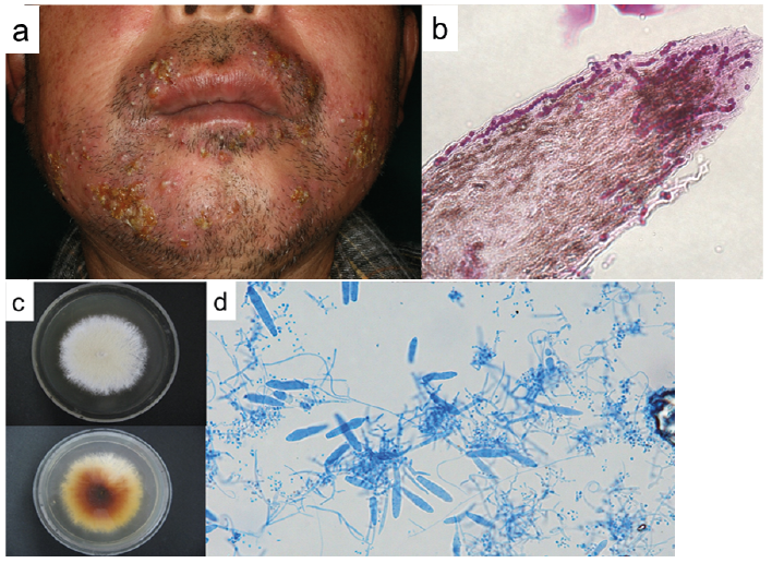
A 69-year-old Japanese man who was a cattle farmer was referred to us with a one-month history of multiple follicular papules and pustules in his beard area. He had been treated with topical and oral antibiotics for 2 weeks before presenting to our hospital. Physical examination revealed multiple follicular papules, multiple pustules with purulent exudate and superficial yellowish crusts, and painful subcutaneous nodules in the beard area. His face was slightly swollen and hairs from the lesion could be plucked easily (Fig. 1a). He was otherwise in a good general health. We performed a biopsy from the cheek lesion and fungal culture from the specimen. Histopathological examination showed a dense perifollicular mixed-cellular inflammatory infiltration with some granulomatous foci in the dermis extending into the subcutaneous tissue. Periodic acid-Schiff (PAS) staining revealed chains of arthro-conidia within and on the hair shafts (Fig. 1b). Culture of the biopsied specimen resulted in cream-white and rapidly growing colonies with a powdery surface (KMU 9922) that developed after 2 weeks of incubation on Sabouraud dextrose agar at 27°C. The reverse side of a colony was yellowish-brown (Fig. 1c). Slide culture of the isolate showed thin-walled macroconidia, spiral hyphae and many grape-like clusters of microconidia (Fig. 1d), suggesting T. mentagrophytes. The isolate KMU 9922 was identified as Arthroderma vanbreuseghemii by sequence analysis of the internal transcribed spacer (ITS) regions of ribosomal DNA genes with 99.8% (643/644) homology to that of the type strain for the fungus previously named A. vanbreuseghemii (RV27960, Gen Bank Accession number AF170452) and were completely identical to T. mentagrophytes under the revised classification (IMF62865, Gen Bank Accession number LC317440)(IMF62865, Gen Bank Accession number LC317440) (Appendix S1). Two weeks after the initial visit, some calves on his farm were found to have multifocal lesions of alopecia with thick crusts on their heads (Fig. 2a, b). An isolate from a crust of one of the calves (KMU 9923) showed identical mycological findings as those of the patient (Fig. 2c, d). The sequence of the isolate from the calf (KMU 9923) showed 100% (644/644) homology to that from the patient (KMU 9922) by sequence analysis of ITS regions of ribosomal DNA genes. Based on these findings, we made a diagnosis of tinea barbae due to T. mentagrophytes infection from the calves. Treatment with oral itraconazole was initiated at a dose of 100 mg/day. Two months later, the lesions resolved and treatment was terminated. However, the lesions recurred one month later and treatment was resumed. The lesions disappeared after another 2 months of treatment with oral itraconazole at a dose of 100 mg/day. At the same time, the patient’s wife was referred to us with a 3-month history of multiple papules and pustules on her pubic area (Fig. 2e). A biopsy specimen revealed perifollicular mixed cellular inflammatory infiltration with focal granulomatous structures and PAS-positive fungal elements within the hair shaft. She did not have any other skin lesions of dermatophytosis and never had direct contact with any of the calves on the farm. Although we failed to detect the pathogen from culture of the biopsy specimen and sequence analysis of the ITS regions of ribosomal DNA genes from a paraffin-embedded specimen, we strongly suspected intrafamilial infection with T. mentagrophytes. The lesions on her pubic area disappeared within 2 months with oral itraconazole treatment. The calves were treated with topical antifungal agents. All the cattle in the farm were born in Japan. There were no other animals, such as dogs and cats, in the farm.
Fig. 1. a) Multiple follicular papules, multiple pustules and painful subcutaneous nodules on the bearded area of the patient. b) Chains of arthroconidia within and on the hair shafts (periodic acid-Schiff stain x400). c) The isolate from the biopsy specimen (KMU 9922). The surface of a colony after 14 days of incubation on Sabouraud dextrose agar at 27°C. d) Slide culture after 14 days of incubation (Cotton-blue stain).
Fig. 2. a) Cattle that the patient had kept. b) Alopecic lesions with thick crusts on the heads of cattle. c) The isolate from the calf (KMU 9923). The surface of a colony after 14 days of incubation on Sabouraud dextrose agar at 27°C. d) Slide culture after 14 days of incubation (Cotton-blue stain). e) Multiple papules and pustules on the pubic area of the patient’s wife.
A. vanbreuseghemii was the designated name of one of the zoophilic teleomorph species of T. mentagrophytes. It was first described by Takashio in 1973 after mating strains were isolated from humans and rodents, such as mice and chinchillas (5). In recent years, a new phylogenetic nomenclature system, based on genetic data, has been proposed (6), and A. vanbreuseghemii previously defined by morphological criteria as a species is now assigned to the newly defined T. mentagrophytes species grouping. According to the literature, isolates formerly reported as “A. vanbreuseghemii” infections of humans are generally contracted from infected pet animals, such as dogs and cats (7, 8). Drouot et al. pointed out that cats with dermatophytoses caused by A. vanbreuseghemii were strictly outdoor cats and a soil source and/or rodent prey were suspected as the main causes of the animal infection (7). In this case, there were neither dogs nor cats in the farm. To the best of our knowledge, A. vanbreuseghemii or, under the new taxonomy, T. mentagrophytes, infection transmission from calves to human skin has not been documented. There was a report of an outbreak of A. vanbreuseghemii infection associated with an infected horse in a veterinary school. It was pointed out in that report that attention must be given to infection transmission from livestock to humans and subsequent transmission from humans to humans (9). Although we failed to identify the pathogen from the skin lesion of the patient’s wife, the risk of intrafamilial infection is a real one. We would like to emphasize that T. mentagrophytes (A. vanbreuseghemii) is transmissible from calves to humans and that simultaneous identification of the pathogenic agent and source of the infection is important in order to prevent recurrence or an outbreak.


 Click to show fullsize
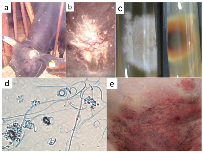 Click to show fullsize