


Department of Pathophysiology and Transplantation, Università degli Studi di Milano, Foundation IRCCS, Cà Granda Ospedale Maggiore Policlinico, Milan, Italy. E-mail: stefano.veraldi@unimi.it
A 68-year-old Caucasian woman was admitted to our department with widespread severe itching. She reported that she was in good general health and that the itching had started approximately 4 months earlier. The patient had been treated unsuccessfully by her general practitioner with diflucortolone valerate cream and oral hydroxyzine.
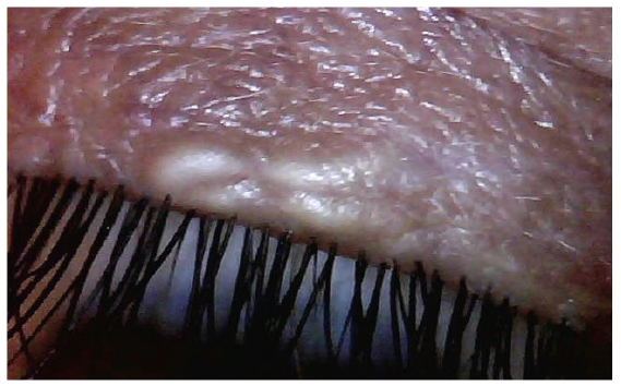
Dermatological examination revealed several lice and nits located on the pubis. Microscopy revealed Pthirus pubis. Furthermore, a cystic-like lesion was observed on the left upper eyelid, with irregular shape, whitish colour, and soft consistency (Fig. 1). The lesion was incised with a scalpel.
What is your diagnosis? See next page for answer.
Fig. 1. A cystic-like lesion, with irregular shape, whitish in colour, located on the left upper eyelid.
Acta Derm Venereol 2019; XX: XX–XX.
Diagnosis: Pthiriasis of the eyelashes
Microscopy revealed several nits on the eyelashes of the left eye (Fig. 2). No lice and/or nits were observed in the eye-lashes of the other eye, although the patient had previously reported itching on all eyelids. It is therefore possible that the infestation had occurred earlier, and not only on the left upper eyelid. Ophthalmological examination was negative. The patient was treated successfully with a foam containing pyrethrins and piperonyl butoxide (1 application/day for 2 consecutive days) on the skin surface, and with petrolatum (3 applications/day for 7 days) on the left eyelid. Follow-up (at 9 months) was negative.
Pthirus pubis Linnaeus, 1758 (Diptera: Anoplura), widely known as crab louse, usually infests the pubis, groin, buttocks, intergluteal fold and perianal region. However, it can also infest, in particular, hairy males or, when the infestation is longstanding, the thighs, abdomen, chest, axillae and beard. Involvement of the scalp is very rare (1).
Pthiriasis of the eyelashes is more common in children, although it is sometimes also observed in adults (2). This pthiriasis is acquired by close contact with infested individuals or contaminated clothing, towels and bedding. It is also considered as a sexually transmitted disease (3). In some cases, pthiriasis of the eyelashes is a manifestation of child abuse (4).
Pthiriasis of the eyelashes usually occurs in both eyelids, although cases with involvement of 1 eye have been reported (5). The eyebrows are rarely involved. This pthiriasis is characterized by erythema, scaling, crusts and lachrymation. Pruritus is the most frequent symptom. Rare symptoms are burning, pain and fever. A common complication is blepharoconjunctivitis (6). Some cases of keratitis have also been reported (7). Bacterial superinfections, with preauricular lymphadenopathy, are very rare.
Numerous therapies have been proposed. Gentle mechanical removal of lice and nits, in spite of the firm adhesion of the nits to eyelashes (8), the application of petrolatum (9) or the application of 0.1–1% yellow oxide of mercury (9), can be considered the most effective and safest treatments. Trimming of the lashes at the base may also be considered (8). Ivermectin is the only oral drug used so far (10). In addition, pillows, bed linen, towels and hats must be washed thoroughly with hot water.
No similar cases of pthiriasis of the eyelashes were found in the literature. It is not known why this cystic-like lesion developed. The patient reported that she repeatedly applied eyeliner, eye shadow and kajal to the eyelids, and this may have been a factor. It is also possible that the lice had infested a tiny infundibular cyst of the eyelashes.
Fig. 2. A nit on an eyelash.


 Click to show fullsize
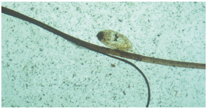 Click to show fullsize