


Department of Pathophysiology and Transplantation, Università degli Studi di Milano - Foundation IRCSS Cà Granda Ospedale Maggiore Poli-clinico, Via Pace 9, Milan, Italy. E-mail: gianluca.nazzaro@gmail.com
A 36-year-old Caucasian woman was admitted to our emergency service because of pruritic cutaneous lesions that had been developing for approximately 3 weeks. She had previously consulted her doctor with a diagnosis of exogenous dermatitis and was treated with a combination of topical corticosteroids and antibiotics.
Dermatological examination revealed red papules, 2–3 mm in diameter, in linear or group configurations on the abdomen and upper extremities. No pustules or vesicles were noted. Some of the papules were excoriated due to the itching. The patient reported that she was living in a flat with her husband and son, neither of whom had cutaneous lesions. No adult mites of Sarcoptes scabiei or fragments thereof or their eggs or scybala were seen under microscopic examination. We confirmed a diagnosis of exogenous dermatitis and recommended examining her entire flat and workplace for the presence of parasites.
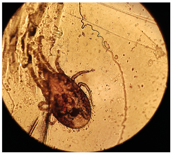
A week later she visited our outpatient service again reporting the same problem. She stated that a professional exterminator had been contracted, but no parasites were found. Her son was now presenting the same dermatitis. The day before she had noted a small “insect” on his right arm and had caught it with adhesive tape. Microscopic examination of the tape solved the case (Fig. 1). Her cat had died recently, and she had bought a hamster.
What is your diagnosis? See next page for answer.
Fig. 1. Microscopy of the adhesive tape provided by the patient.
Acta Derm Venereol 2019; XX: XX–XX.
Diagnosis: Tropical rat mite (Ornithonyssus bacoti) dermatitis
The tropical rat mite, Ornithonyssus bacoti, is an obligate blood-feeding ectoparasite belonging to the family Macronyssidae, which is found mostly in wild rats from tropical and moderate climate zones (1). In case the rodents are not available, human beings may become the victim of this haematophagous mite (2). In 1913, the first case of rat mite dermatitis was reported in Australia (3). In the USA, it was first reported in 1923. In Europe, O. bacoti was first observed in Germany, probably carried across the oceans by ship on infested rats or mice (4).
This dermatitis is not distinctive, usually consisting of small erythematous papules, either singular or located in groups. The definitive diagnosis of this ectoparasitosis requires the detection of the parasite. The patient is usually unaware of the cause because tropical rat mites are active at night and seek dark hiding places during daytime. Although the blood meal in the skin lasts at most 20 min and happens at night, the mites usually cannot be detected on the skin (3, 5). The rat mite is not known to be the vector of any infectious diseases in humans; however, some authors have reported the tropical mite as a vector of human typhus (6, 7). Prompt identification of the rat mites and the eradication of the parasites with adequate acaricides, such as permethrin, can prevent the disease spreading.
O. bacoti has 5 developmental stages: egg, larva, protonymph, deutonymph and adult. After a blood meal, the pregnant female drops from the host and lays up to 9 eggs at a time, which hatch into non-feeding larvae in 1.5 days. The entire life cycle can take as little as 11–13 days under optimum conditions (8). Tropical rat mites are much more hairy than other mite species. They are frequently confused with bird mites (Dermanyssus gallinae) or the Nordic bird mites (Ornithonyssus sylviarum), which also belong to the Macronyssidae and have similar morphological characteristics. Certain morphological structures (e.g. hairiness, caudally pointed scutum (dorsal plate), typical form of the anal plate with a cranial anus) allow for differentiation of the tropical rat mite from other mite species (9).
No epidemiological studies determining the prevalence of rodent mite dermatitis have been published. D’Ovidio et al. (10) indicate that the prevalence of O. bacoti in exotic pet mammals in an area in southern Italy is 9.8%, suggesting that the occurrence of this mite is currently underestimated. In our opinion, the true incidence of the disease is greater than suggested by the low number of case reports presents in human and veterinary literature, possibly due to the difficulty of detection and accurate identification of this ectoparasite.


 Click to show fullsize