


Department of Dermatology, The University of Tokyo Graduate School of Medicine, 7-3-1, Hongo, Bunkyo-ku, Tokyo 1138655, Japan. *E-mail: koukinakamura-tky@umin.ac.jp
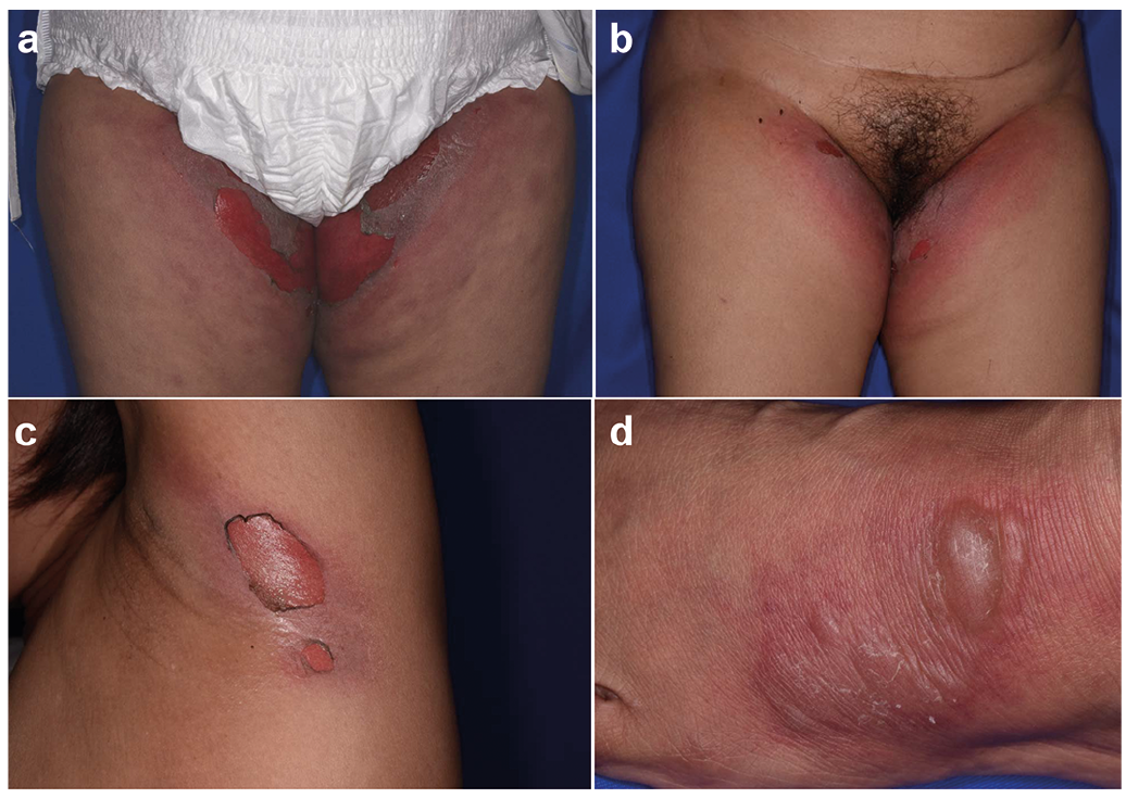
A 54-year-old Burmese woman presented to our dermatology clinic with a 2-day history of blisters and skin erosions surrounded by pruritic erythema in her inguinal region (Fig. 1a). She had experienced a similar rash twice on the same sites, 1 month and 1 year earlier (Fig. 1b). In this third episode, the patient had more extensive skin involvement compared with the previous episodes: the rash appeared not only in the inguinal lesion, but also on her neck, axillae (Fig. 1c), and feet (Fig. 1d). No mucosal eruption was seen in the oral cavity or genital area. For all episodes the patient had taken loxoprofen tablets 1 or 2 weeks earlier for her back pain. A thorough review did not find any other drugs, herbs, or use of supplements. The patient was afebrile, and her general status was normal. Laboratory tests revealed a white blood cell count of 11,200 /mm3 with 79% neutrophils and a C-reactive protein level of 123.4 mg/l. A skin biopsy demonstrated subepidermal blisters and vacuolar interface dermatitis with perivascular infiltration of eosinophils, lymphocytes, and neutrophils. In the epidermis, there were scattered dyskeratotic keratinocytes (Fig. 2). Loxoprofen was stopped and treatment of the rash with topical cortico-steroids was started, which resolved the skin lesions leaving residual hyperpigmentation. The patient has been free from further recurrence since stopping loxoprofen.
What is your diagnosis? See next page for answer.
Fig. 1. Clinical features of the rash. (a) Skin erosion surrounded by pruritic erythema in the patient’s inguinal region. (b) Similar rash in the identical site in the previous episode. (c) Skin erosion in her left axilla. (d) Blisters and infiltrated erythema on the dorsum of her left foot.
Fig. 2. Histopathological features of the rash. (a) Subepidermal blisters and vacuolar interface dermatitis are observed. (b) Scattered dyskeratotic keratinocytes in the epidermis and perivascular infiltration of eosinophils, lymphocytes, and neutrophils in the dermis (haematoxylin-eosin stain: a: 100×, b: 200×).
Acta Derm Venereol 2019; XX: XX–XX.
Diagnosis: Generalized bullous fixed drug eruption
Generalized bullous fixed drug eruption (GBFDE) is a rare variant of drug eruption, characterized by symmetrical widespread blisters with a peripheral rim of erythema, which appears recurrently on identical sites. The development of GBFDE is sometimes preceded by milder episodes of fixed drug eruption, while other cases emerge de novo (1). The rash typically appears on the extremities and the trunk, but is less likely to invade the face or head (2). Two patterns of the rash have been reported: one with oval or egg-shaped lesions, and one with well-demarcated, but not oval, lesions. Although in the second form the demarcation may not be obvious at first, both types eventually form a sharp erythematous margin. Histopathology revealed vacuolar interface dermatitis and subepidermal blisters with eosinophilic infiltration, as seen in other types of drug eruption (3). The common causative agents of GBFDE include non-steroidal anti-inflammatory drugs (NSAIDs), antibiotics, metamizole, and sulphonamides (1, 2, 4). When the patient’s medical history does not reveal a causal relationship between the rash and drugs, patch-testing at the involved sites can detect it accurately in approximately one-third of cases (2). GBFDE is often misdiagnosed as other subtypes of drug eruption, such as Stevens-Johnson syndrome (SJS) and toxic epidermal necrolysis (TEN) (5, 6). The prominent features of GBFDE compared with SJS/TEN include older age, less mucosal involvement, and less systemic symptoms, such as malaise and fever (2). Immunohistological comparison demonstrated infiltration of more CD4+ cells, more FoxP3+ cells, less CD56+ cells, and less granulysin+ cells in GBFDE. In addition, the serum level of granulysin, a key molecule in SJS/TEN keratinocyte death, was lower in GBFDE than in SJS/TEN (3). These insights indicate that FoxP3+ regulatory T lym-phocytes suppress granulysin production by CD8+ cytotoxic T lymphocytes and CD56+ natural killer cells in GBFDE (7), which highlights the difference in pathogenesis between GBFDE and SJS/TEN. The mainstay of treatment is cessation of the causative agents and supportive care. In general, GBFDE has better prognosis and can be managed by much more conservative treatment than SJS/TEN (6). However, a recent case-control study reported that mortality in GBFDE was comparable with that of SJS/TEN (22% vs. 28%) (8). Clinicians should therefore keep GBFDE in mind as a potentially fatal adverse drug reaction.
The authors have no conflicts of interest to declare.


 Click to show fullsize
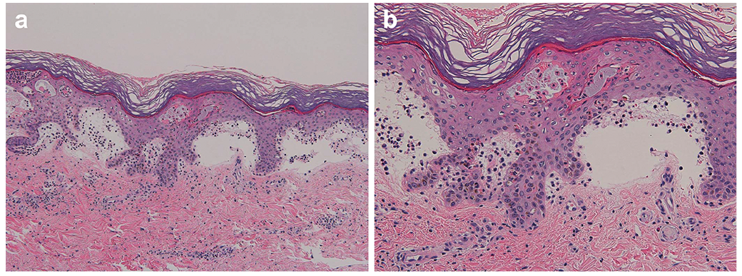 Click to show fullsize