


1STD Institute and 3Department of Dermatopathology, Shanghai Dermatology Hospital, Tongji University School of Medicine, Shanghai 210043, and 2Institute of Dermatology, Chinese Academy of Medical Sciences and Peking Union Medical College, Nanjing, China. *E-mails: md_longfuquan@163.com or zpyls@yahoo.com
Accepted Aug 22, 2019; E-published Aug 22, 2019
Syphilis is a chronic sexually transmitted infection caused by Treponema pallidum. The protean cutaneous manifestations of secondary syphilis span a wide spectrum and have earned the name of “the great imitator” (1, 2). Secondary syphilitic lesions usually occur within 3 months after initial exposure to T. pallidum and last for 4 to 12 weeks (3). However, co-infection with HIV may alter the clinical presentation and course (4). Here, we report a long-term misdiagnosed HIV-seropositive homosexual man presenting with psoriasis-like secondary syphilitic lesions, which appeared approximately 3 years after initial infection and had a protracted course over one year. Such notable deviations from the normal expectations have never been reported.
A 30-year-old man who complained of skin rashes and hair loss for more than one year was referred to our STD clinic in August 2017. He denied any sexual behaviour except for two episodes of unprotected anal sex with men in July 2012 and March 2013. His serological tests for HIV and syphilis were all negative before his last sexual encounter. He was diagnosed with HIV infection in July 2013; at that time his CD4+ T-cell count was 46 cells/ul. He concealed his HIV status from his family members and doctors. He started highly active antiretroviral therapy in September 2016, but he ceased treatment voluntarily after 3 months because of profoundly pessimistic thoughts. The patient had experienced gradual hair loss and recurrent scalp dermatitis since July 2016. He had been diagnosed with psoriasis with concomitant alopecia for more than one year and was tested for syphilis and HIV in the dermatology clinics. He received intermittent therapy with a topical steroid ointment without improvement. The lesions progressively expanded to involve his neck, shoulders, wrists, feet and genitals.
Physical examination revealed patchy alopecia and multiple varisized infiltrated flat red psoriasis-like plaques involving the scalp, neck, face, shoulders, feet and genitals (Fig. 1). He had axilla, neck, submaxilla and groin lymphadenopathy. The T. pallidum particle assay was positive with a toluidine red unheated serum test titre of 1:64. His HIV viral load was 151 copies/ml, and his CD4+ T-cell count was 15 cells/µl. Cerebrospinal fluid tests indicated slightly elevated leukocyte (15 cells/μl) and protein levels (60 mg/dl), but the Venereal Disease Research Laboratory test was negative. A skin biopsy taken from a left wrist lesion revealed that the epidermis was irregularly proliferated, dermal papillae were oedematous, and perivascular plasma cells had infiltrated the superficial dermis (Fig. 2a). Immunohistochemical analysis using a polyclonal antibody against T. pallidum showed numerous spirochetes (Fig. 2b). The diagnosis of secondary syphilis and HIV infection was confirmed. The patient received 4 million units of benzylpenicillin intravenously every 4 h for 14 days. His skin lesions and patchy alopecia improved significantly 6 weeks later (Fig. 2c, d). The patient is still being followed.
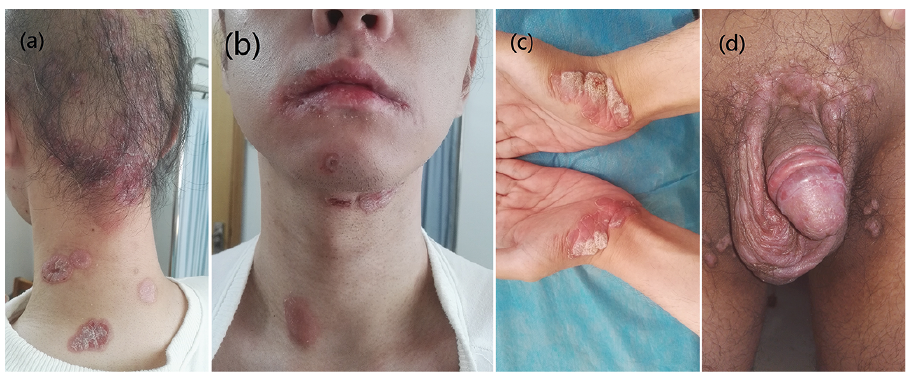
Fig. 1. a) Patchy alopecia and red psoriasis-like plaques on the patient’s scalp and neck. b) Red plaques on the patient’s neck and jaw and dry erythema on his lips. Several varisized red and flat-topped, hypertrophic, verrucous and psoriasis-like plaques on the patient’s wrists (c) and genital area (d).
Fig. 2. A skin biopsy specimen from the patient’s left wrist lesion revealed that the epidermis was irregularly proliferated, dermal papillae were oedematous, and perivascular plasma cells infiltrated in the superficial dermis (haematoxylin-eosin stain, original magnification ×100) (a). Immunohistochemical analysis using a polyclonal antibody against T. pallidum showed numerous spirochetes (original magnification ×400) (b). Six weeks after treatment, the lesions on the patient’s scalp (c) and wrists (c) almost disappeared.
Syphilis is one of the most important and neglected diseases. A resurgence in syphilis has been observed in many parts of the world in recent decades, especially in China (2, 5). The alarming resurgence has been characterized by high rates of concomitant HIV infection, especially among men who have sex with men (MSM) (6). Co-infection with HIV affects the initial presentation, disease course, diagnosis, and treatment of syphilis (7, 8). It has been documented that HIV often accelerates the progression of syphilis (4, 6, 9). However, our patient did not develop cutaneous eruptions of secondary syphilis until approximately 3 years after T. pallidum infection (we cannot be sure of the precise infection date based on the patient’s declarations), and had a protracted course of more than one year. Similarly, Carnauba et al. (10) reported that a 44-year-old woman suffering from secondary syphilis and coinfected with human T-lymphotropic virus-1, a causative agent that, like HIV, predominately invades CD4+ T cells; it presents with syphilitic lesions with a two-year delayed-onset and lasts for 6 months.
Previous studies have indicated that the clinical course and manifestation of secondary syphilis may be determined by the deposition of circulating immune complexes in highly susceptible tissue and influenced by the balance between delayed-type hypersensitivity and humoural immunity (11, 12). The significantly reduced CD4+ T-cell count in our patient may have played a role in the delayed onset of his symptoms, suggesting that cellular immunity can modify the evolution of the infection (13, 14). However, the exact mechanism of how HIV and T. pallidum co-interact with the host is still unclear.
The early diagnosis and treatment of syphilis in HIV-infected patients is challenging due to atypical clinical features and deviations from the expected course (15). In this case, the patient had been misdiagnosed with psoriasis for more than one year and was finally diagnosed by skin lesion biopsy and serological tests for syphilis. Such unusual presentations should remind clinicians to be alert to the atypical clinical manifestation of syphilis, especially in patients infected with HIV.
The authors have to no conflicts of interest declare.


 Click to show fullsize
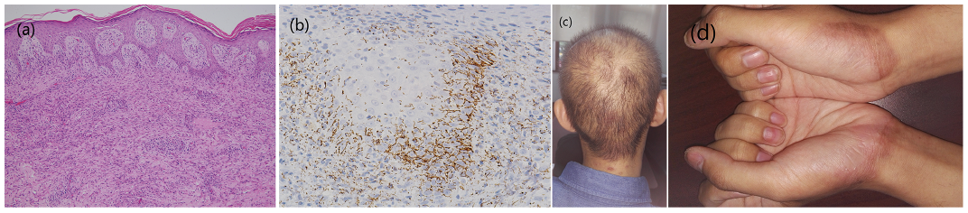 Click to show fullsize