


1Section of Dermatology, DISSAL, University of Genoa, and San Martino Policlinic Hospital, Genoa, 2Laboratory of Molecular and Cell Biology, IDI-IRCCS, Rome, 3Department of Clinical-Surgical, Diagnostic, and Pediatric Science, Institute of Dermatology, Fondazione IRCCS Policlinico San Matteo, Pavia, 4Section of Dermatology, Department of Health Sciences, University of Florence, Florence, 5UCO of Dermatology, Fondazione IRCCS Ca’ Granda Ospedale Maggiore Policlinico, 6Department of Physiopathology and Transplantation, Università degli Studi di Milano, 7UOC of Pediatric Dermatology, Fondazione IRCCS Ca’ Granda Ospedale Maggiore Policlinico, Milan, 8Dermatology Unit, Department of Surgery and Intensive Care, IRCCS G. Gaslini Children’s Hospital, Genoa, and 9Institute of Dermatology, Fondazione Policlinico Universitario A. Gemelli IRCCS, Università Cattolica del Sacro Cuore, Rome, Italy
#Both authors contributed equally to this work.
Linear immunoglobulin A (IgA) bullous dermatosis (LABD) is characterized by the presence of multiple IgA autoantibodies, and a comparatively lesser number of IgG antibodies, directed against different hemidesmosomal antigens. The main autoantigens are LAD-1, LABD-97, BP180 and BP230, type VII collagen and laminin 332. The serology of 54 Italian patients with LABD was studied retrospectively using enzyme-linked immunosorbent assay (ELISA), immunoblotting assay, and indirect immunofluorescence on monkey oesophagus and salt-split skin. Among these, indirect immunofluorescence of salt-split skin elicits the greatest sensitivity. Sixty-three percent of the sera were observed to be positive, with a lamina lucida pattern observed in 48%, a sub-lamina densa pattern in 2% and a mixed pattern in 13% of cases. IgA reactivity to LAD-1 on immunoblotting was found in 52% of sera, to BP180-NC16A by ELISA in 32% and to BP230 in 26%. Only 17% of patients had circulating IgG autoantibodies. LAD-1 was determined to be a major autoantigen of the lamina lucida subtype. Combined serological assays demonstrated a high sensitivity (82%), suggesting that this approach could support diagnosis when a biopsy is not feasible or direct immunofluorescence results are negative.
Key words: linear IgA bullous disease; humoral immune response; LAD-1; BP180; BP230; diagnostic sensitivity.
Accepted Jan 9, 2020; Epub ahead of print Feb 3, 2020
Acta Derm Venereol 2020; 100: adv00070.
Corr: Emanuele Cozzani, Section of Dermatology, DISSAL, University of Genoa, Via Pastore 1, 16132, Genoa, Italy. E-mail: emanuele.cozzani@unige.it
Linear IgA bullous dermatosis is a rare autoimmune disease, marked by blistering, affecting both children and adults. It is characterized by auto-antibodies targeting various types of skin proteins. We retrospectively studied 54 Italian patients with this disease. The auto-antibody targets were investigated using various blood tests (enzyme-linked immunosorbent assay (ELISA), immunoblotting and indirect immunofluorescence on monkey oesophagus and salt-split skin). Indirect immunofluorescence on salt-split skin elicited the greatest diagnostic sensitivity. The most common target was LAD-1, followed by BP180-NC16A and BP230. Eighty-two percent of patients had circulating IgAs, and only 15% possessed circulating IgG auto-antibodies. Combined serological tests supported the diagnosis of linear IgA bullous disease.
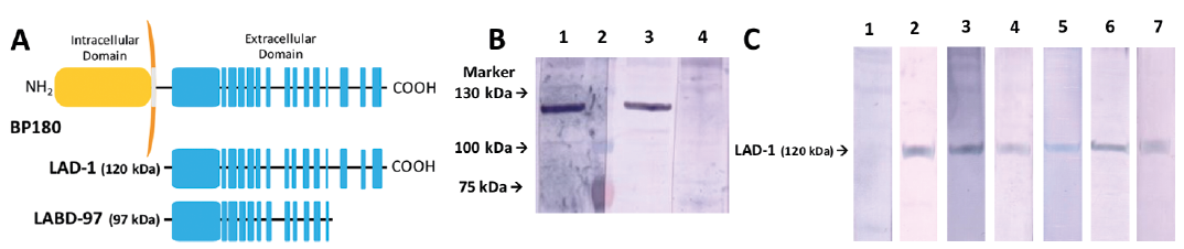
Linear IgA bullous dermatosis (LABD) is an IgA-mediated autoimmune bullous disorder occurring in adults and children, characterized by sub-epidermal blisters and linear deposits of IgA at the basement membrane zone (BMZ) (1). Compared with other immunobullous diseases, LABD is immunologically a heterogeneous disease with pathogenic IgA autoantibodies against different hemidesmosomal antigens (2). Researchers have identified multiple targets over years of research, including collagen VII (3–5), laminin 332 (6), an unidentified 285-kDa protein (7), bullous pemphigoid 230 kDa antigen (BP230) (8), bullous pemphigoid 180 kDa antigen (BP180) (collagen XVII) (8), a 120-kDa protein (linear IgA disease-1 antigen; LAD-1) (9–11) and a 97-kDa protein (LABD-97) (12–14) (Fig. 1A). The last 2 antigens were subsequently denoted as parts of the extracellular domain of BP180 ectodomain (10, 15–18).
Fig. 1. (A) Schematic representation of human BP180 and its fragments LAD-1 and linear IgA bullous dermatosis (LABD)-97. (B) The shed ectodomain of BP180 (LAD-1) is recognized by IgG and IgA autoantibodies as a 120-kDa polypeptide. Anti-BP180-positive bullous pemphigoid serum bind 120-kDa polypeptide by IgG (lane 1), molecular weight marker (lane 2), an anti-BP180 positive LABD serum bind 120-kDa polypeptide by IgA (lane 3), healthy volunteer serum does not bind 120-kDa polypeptide by IgA (lane 4); (C) IgA reactivity to LAD-1 (120 kDa) detected by immunoblotting: lane 1, healthy volunteer serum; lane 2, LABD control serum; lanes 3–7, LABD patient sera from the present study.
BP180 is a hemidesmosomal type II transmembrane protein comprising an intracytoplasmic domain (NC16C domain), a transmembrane domain (NC16B domain), and an extracellular region with 15 collagenous domains and 16 non-collagenous domains (NC16A to NC1) (Fig. 1A) (19). The NC16A domain is the main epitope in bullous pemphigoid. BP180 can be cleaved physiologically within the NC16A domain by ADAM9/17 (16, 20) or pathologically by plasmin (21) and neutrophil elastase (22), into a 120-kDa extracellular fragment corresponding to LAD-1 (Fig. 1B). Further processing of LAD-1 within the carboxyl-terminal NC4 domain yields a 97-kDa fragment corresponding to LABD-97 (23, 24).
LABD differs from bullous pemphigoid, in which IgG autoantibodies preferentially target other parts of BP180 (25, 26). Furthermore, it has been shown that IgA autoantibodies from subsets of patients with LABD recognize epitopes within the NC16A region (27, 28).
LABD displays 2 major binding patterns of IgA autoantibodies in indirect immunofluorescence of salt-split skin (IIF-SSS) and in immuno-electron microscopy. The majority of the LABD sera IgA react with the epidermal side of IIF-SSS, corresponding to the lamina lucida type sera, while a fewer number of IgA react with the dermal side of IIF-SSS, corresponding to the sub-lamina densa type sera (29, 30). A limited number of IgA elicit mixed reactivity to both locations. The type VII collagen in anchoring fibrils was determined to be the primary target of IgA antibodies from patients with sub-lamina densa type of LABD (4, 5, 31). In contrast, most patients with lamina lucida type LABD have IgA autoantibodies targeting LABD-97 (12–14) and LAD-1 (9, 10).
The autoimmune heterogeneity considers both targeted antigens as well as the involved Ig class. Apart from auto-reactive IgAs, IgGs were also reported to be associated with LABD, which are detected as linear deposits at the BMZ in direct immunofluorescence (DIF) and detected in patients’ sera in IB testing or by enzyme-linked immunosorbent assay (ELISA) (6).
The present retrospective study characterized the humoral immune response of 54 Italian patients with LABD using standard approaches, such as IIF-SSS and commercial and adapted ELISAs. In addition, this study suggests that LAD-1 is the major antigen LAD-1 as a major auto-antigen in Caucasian patients affected by the lamina lucida type LABD.
Patients
Sera were collected from 54 patients with LABD, including children and adults from different Italian clinical centres (Milan, Pavia, Genoa, Florence and Rome) between 2012 and 2017, for retrospective study. All patients typically demonstrated clinical, histopathological and immunopathological features of LABD (32): (i) vesicular or bullous eruption involving skin and mucous membranes; (ii) sub-epidermal blisters infiltrated predominantly by neutrophils detected in lesion biopsies; (iii) linear IgA-deposit pattern at the BMZ detected by DIF. Patients with both IgA and IgG deposits at the BMZ detected by DIF were excluded. Due to the retrospective design of the study, information on drug-induce LABDs was not available for all patients.
Indirect immunofluorescence
IIF was performed on monkey oesophagus (MO) samples using commercial slides (BioSystems S.A. Barcelona, Spain). IIF-SSS was performed using a commercial kit (Immco Diagnostics, NY, USA). All sera were diluted in the ration 1:20 with phosphate-buffered saline (PBS). The slides were incubated with the diluted sera for 30 min. After washing in PBS, the slides were coated with fluorescein isothiocyanate-conjugated goat anti-human IgA or IgG (Kallestad Diagnostic, Chaska, MN, USA) for 30 min. Following a further PBS wash, the slides were mounted in buffered glycerine and examined under a fluorescence microscope (Leitz, Wetzlar, Germany). An IIF titre greater than 1:40 was considered positive.
Enzyme-linked immunosorbent assay for IgG and IgA detection
LABD IgG autoantibodies against BP180-NC16A and BP230 were characterized using commercial ELISA kit (MBL Co, Nagoya, Japan), according to the manufacturer’s protocol. To compare the results obtained from different plates, optical densities (ODs) of the test samples were adjusted according to positive and negative control samples supplied in each kit. The protocol had been modified to an extent for the detection of IgA autoantibodies against BP180 and BP230. Briefly, BP180 and BP230 microwell strips were incubated with LABD patient sera diluted 50 times and appropriate control sera. After washing, the bound antibodies were detected using 10.000-fold diluted HRP-conjugated anti-human IgA antibody (Sigma-Aldrich, St Louis, MO, USA). Colour development was achieved using 3,3’,5,5’-tetramethylbenzidine (Sigma) and after the reaction ceased, OD was measured at 450 nm with the correlation wavelength set at 620 nm using a microplate reader (Bio-Rad Laboratories Inc., Hercules, CA, USA). The cut-off value was determined to be the mean OD measured in a cohort of serum samples collected from 30 healthy volunteers and 3 standard deviations.
LAD-1 preparation and immunoblotting analysis
LAD-1, the 120-KDa soluble ectodomain of BP180, was isolated from the culture medium of normal keratinocytes derived from human epidermis). Keratinocytes were grown at confluence and shed ectodomain of BP180 released in the medium was precipitated by the addition of ammonium sulphate (30% saturated solution) and incubated overnight at 4°C. Precipitates were collected, re-suspended in 25 mmol/l Tris-HCL, pH 7.8, 65 mmol/l NaCl and 5 mmol/LEDTA and dialysed against the same buffer overnight at 4°C. LAD-1 was electrophoresed on a 6% polyacrylamide gel in reducing conditions.
Immunoreactivity was detected after transfer to polyvinylidene difluoride membrane (Immobilon-P, Millipore Corporation, Billerica, MA, USA), by incubation with a 1:20 dilution of patient or control sera, followed by incubation with alkaline phosphatase-labelled secondary antibodies against IgA.
Study population
Serum samples were collected from 54 patients with LABD (12 children and 42 adults), 30 females and 24 males, with overall mean age 41 ± 31 years (age range 1–93 years).
Indirect immunofluorescence results
Presence of IgA at the BMZ was detected in 8 out of 54 (15%) serum samples from LABD patients, as tested by IIF on MO, while 34/54 (63%) serum samples from LABD patients were IgA-positive, as tested by IIF-SSS on human skin (Table I). In particular, 26/54 (48%), 1/54 (2%) and 7/54 (13%) serum samples were determined to have lamina lucida, sub-lamina densa and mixed patterns, respectively (Table I). Considering adults and children individually: serum samples collected from 6/12 (50%) children and 28/42 (67%) adults reacted with IIF-SSS.
With regards to IgG, none of the serum samples reacted with IIF on MO, while 1/54 (2%) serum samples (which was extracted from an adult) reacted to the epidermal side of IIF-SSS on human skin.
Table I. Immunological test results in 54 patients with linear IgA bullous dermatosis (LABD)
IgA reactivity profile
Overall, IB results indicated IgA reactivity to LAD-1 in 28/54 serum samples (52%) (Table I, Fig. 1C). Anti-LAD-1 IgA were detected by IB in 8/12 (67%) serum samples from children and from 20/42 (48%) serum samples adults. BP180-NC16A ELISA results were positive in 17/54 patients (32%) and BP230 ELISA results were positive in 14/54 patients (26%) (Table I).
Considering the IIF-SSS pattern groups, serum samples from patients with lamina lucida binding pattern had variable reactivity to different epidermal antigens, as shown in Table II. Some of the sera samples reacted with multiple epidermal antigens (Table II). Altogether, IgA from 24/26 (92%) samples binding to lamina lucida reacted with at least 1 of the epidermal antigens studied.
Table II. IgA reactivity profile in 34 indirect immunofluorescence- salt-split skin IgA-positive patients
IgG reactivity profile
ELISA with BP180-NC16A and BP230 were positive for 3/54 (6%) and 5/54 patients (9%), respectively (Table I). Overall, circulating IgG antibodies anti-BP180-NC16A and/or anti-BP230 detected by ELISA were detected in 9/54 (17%) serum samples, while the total number of serum samples with IgG autoantibodies detected either by DIF, IIF-SSS or ELISA were 7/54 (13%).
These results confirm the heterogeneity of IgA auto-antibody (autoAbs) targets in LABD. The predominant Ig class auto-antibody in the present cohort of patients with LABD was IgA, whereas IgG was present in only 22% of patients. Auto-reactive IgAs in serum samples primarily targeted LAD-1, while targeting BP180-NC16A and BP230 to a lesser extent.
The processed extracellular part of BP180 represents the primary antigenic stimulus in LABD, indicating that the processing induces neo-epitope formation (33). Extracellular regions of BP180 produced by removal of intra-cytoplasmic and transmembrane domains were reported to be recognized by 83% of LABD serum samples (26). Schumann et al. demonstrated that IgA targeted the soluble ectodomain more efficiently than the full-length protein in patients with LABD; 50% of the adult LABD serum samples and 100% of child LABD serum samples recognized this target (11). Likewise, in our experiments, 48% of adult LABD serum samples and 67% of child LABD serum samples reacted with LAD-1. In line with previous study, serum samples from children with LABD seem to particularly react with LAD-1 in comparison with adults; this finding could suggest a major role of neo-epitopes present in the LAD-1 antigen in the disease pathogenesis in children. However, due to the low number of patients in our cohort (8 patients) and in the cohort from Schumann’s study (7 patients), no conclusion can be drawn. This issue should be investigated further on a larger cohort. In the present study, approximately 50% of the patients were positive for anti-LAD-1 IgA, and this proportion increased to 73% when only lamina lucida type patients were considered. Moreover, there was a strong correlation (90%) between lamina lucida positive sera and reactivity to BP180 (both LAD-1 and NC16A) and BP230. IgA positive against BP180-NC16A was detected consistently in a significant number (38%) of patients, which is in line with previous reports (6, 27, 28, 34). Notably, only 50% of these patients had targeted both LAD-1 and BP180-NC16A, while 50% exclusively targeted BP180-NC16A. Also, as anticipated, most patients positive for LAD-1 IgA (detected by IB) were negative for BP180-NC16A IgA (detected by ELISA). Notably, autoAbs directed against BP230 were more frequently detected in patients with LABD when BP180 autoAbs were also present. This association could depend on an intermolecular epitope spreading phenomenon (35, 36). Specifically, the pathological progression could induce damage in epithelial cells and result in abnormal exposure of the intracellular BP230, inducing the production of autoAbs (35). Undoubtedly, IB for 285 kDa antigen, LABD-97, full-length BP180 and type VII collagen might have yielded significant results; however, due to economic and technical limitations these tests were not conducted.
The concentration of circulating IgGs reactive to BMZ antigens was significantly low (17%). This is in sharp contrast with the results reported recently by Ohata et al., in which positive circulating IgG autoantibodies were detected either by IIF, IB or ELISA in 53% of their serum samples (6). The current study probably observed lower rates of auto-reactive circulating IgGs, as unlike the protocol adopted by Ohata et al., we did not test for IgGs directed against a wider variety of autoantigens. Considering LABD and BP to be opposite ends of the same spectrum, as suggested by Ohata et al. (6), might be inaccurate; however, these 2 diseases are overlap significantly in terms of clinical and serological aspects (37).
The higher positivity rates of IIF-SSS compared with IIF on MO confirmed the higher sensitivity of IIF-SSS in detecting auto-reactive IgAs. Our results confirm that the lamina lucida type is the most frequent variant of LABD. This is in line with a previous report suggesting that IgA reactivity is more prevalent on the epidermal end than on the dermal end of IIF-SSS (6).
The present study denotes that serological diagnosis of LABD patients can be performed using different assays for detection of IgG and IgA circulating autoantibodies (IIF, IB on LAD-1, BP180 and BP230 ELISAs). Notably, the combined sensitivity allows detection of auto-antibodies in 83% of patients (45/54). Specifically, the most efficient process hierarchy involves DIF, followed by IIF-SSS as the most sensitive approach, followed by IB on LAD-1 and ELISA on IgA BP180, and IgA BP230 ELISA performed in case of negative results (which in one case was the only positive serological test). Notably, none of the LABD serum samples that resulted negative for circulating IgA antibodies in IIF, IB for LAD-1 or ELISA for BP180, were positive for IgG ELISAs.
This study has certain limitations; it focused specifically on the epidermal antigens targeted in LADB, and only a limited number of paediatric patients were studied.
Conclusion
LABD is characterized by a heterogeneous humoral response, largely comprising IgA auto-antibodies and a lesser number of IgG autoantibodies. It is yet to be established if these auto-antibodies elicit pathogenicity. Several antigens of the BMZ are targeted by auto-antibodies in LABD. LAD-1 is a major auto-antigen of the lamina lucida subtype, particularly in children. DIF remains the most preferred diagnostic method for detection of LABD. However, the combination of several serological assays could an important approach to support diagnosis, especially in cases in which a biopsy is not feasible or the DIF results are negative.


 Click to show fullsize
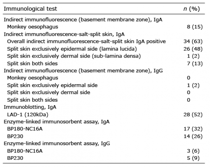 Click to show fullsize
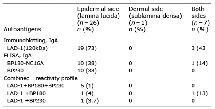 Click to show fullsize