


1Dr. Phillip Frost Department of Dermatology and Cutaneous Surgery and Miami Itch Center, Miller School of Medicine, University of Miami, Miami, USA, 2Institute of Medical Psychology, Justus-Liebig-University, Giessen, Germany, 3Department of Dermatology, Lewis Katz School of Medicine, Temple University, Philadelphia, and 4Department of Pathology, School of Medicine, University of Virginia, Charlottesville, USA
Accepted Feb 19, 2020; Epub ahead of print Mar 4, 2020
Acta Derm Venereol 2020; 100: adv00129
Psychological stress has been shown to be related to an aggravation of atopic dermatitis (AD) (1). Therefore, addressing the role of stress in the pathology of AD is of prime importance. The hypothalamic-pituitary-adrenal (HPA) axis and sympathetic-adrenal-medullary (SAM) axis are major descending stress-response pathways from the hypothalamus that regulate a variety of physiological functions, including immune and skin barrier functions (1). Previous studies have demonstrated that the HPA and SAM axes are important physiological systems that react in the presence of stress. Usually, cortisol release is increased by acute stress. However, the cortisol response to stress has been shown to be blunted in AD patients, which in turn facilitates the Th2 dominance and hyperproduction of IgE, and affects skin barrier functions (1–4). These changes due to stress are considered to be the major factors to aggravate AD. However, it is still unknown how stress affects HPA and SAM functions. The hypothalamus is a brain structure that plays a pivotal role in the neuroendocrine aspect of stress. Previous studies have demonstrated that anatomical changes in the hypothalamus are related to dysfunctions of the HPA axes (5, 6). It is possible that stress affects hypothalamic volume, as a recent study in patients with anxiety disorder demonstrated a significant reduction of hypothalamic volume (7). However, as of yet, no study has explored the relationship between stress and hypothalamic volume in AD patients. This pilot study addressed this question using Voxel-Based Morphometry (VBM) in an explorative manner.
We collected brain anatomy images and stress scores from 14 AD patients (mean age: 32 ± 11 years old, male/female: 5/9) and 11 healthy subjects (age: 30 ± 8 years old, male/female: 4/7). From some of the subjects, we obtained further data in addition to the anatomical measurement (also see 8). The images were acquired on a Siemens Verio 3T MR scanner at Temple University hospital. Stress level a week prior to the MRI measurement day was assessed using the Perceived Stress Questionnaire (PSQ; 9). All participants signed an informed consent approved by the Institutional Review Board of the Temple University School of Medicine. Individual MRI images were processed with statistical parametric mapping (SPM8) (http://www.fil.ion.ucl.ac.uk/spm) and the VBM8 toolbox (http://dbm.neuro.uni-jena.de/vbm.html) to obtain the gray matter (GM) whole brain images in individual subjects. These images were nonlinearly normalized to the brain template supplied with the VBM 8 toolbox. This process preserved the local gray matter volume as identified in native space corrected for total brain size. Prior to statistical analysis, the spatially normalized GM images were smoothed with an 8 × 8 × 8 mm3 FWHM kernel.
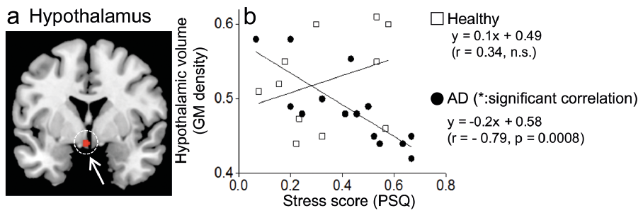
We conducted a correlation analysis using GM images and stress scores obtained from 14 AD patients with focusing on the hypothalamus. First, we applied an uncorrected p < 0.001 at the voxel level. Then, a small volume correction (SVC) using a 10 mm sphere was applied. The center of sphere ((x, y, z) = (2, –1, –12)) was determined based on a previous study which identified the MNI coordinate corresponding to the paraventricular nucleus (PVN) of the hypothalamus, a key nucleus to regulate the HPA and SAM (10). The sphere encompassed the bilateral hypothalamus. Statistical threshold was set at p < 0.05 (Family-Wise Error (FWE)). Confounding factors such as disease duration and age were covariated out in this analysis. The same analysis was conducted using GM images obtained from 11 healthy subjects. As shown in Fig. 1a and b, hypothalamic GM density was significantly negatively correlated with perceived stress in AD patients, but not in healthy subjects. Here, a non-significant positive correlation occurred for healthy subjects. The mean stress score was 0.42 ± 0.19 in AD patients and 0.34 ± 0.18 in healthy controls (possible range: 0–1). There was no significant difference between the two groups in perceived stress [AD: 0.42 ± 0.19, healthy: 0.34 ± 0.18, T (23) = 1.1, p = 0.28)] or GM density [AD: 0.49 ± 0.05, healthy: 0.52 ± 0.06, T (23) = –1.57, p = 0.13].
Fig. 1. Gray matter (GM) density in the hypothalamus. (a) A location in the hypothalamus where GM density was significantly negatively correlated with stress score (red dot and white arrow) was superimposed on the MNI brain template (https://www.nitrc.org/projects/mricron). (b) A scatter plot of GM density in the hypothalamus and stress score. We extracted mean GM density from the hypothalamus using Marsbar (http://marsbar.source-forge.net/). In detail, a region of interest (ROI) was placed on the hypothalamus. The ROI was a sphere with 3 mm radius centered on the MNI coordinate that showed a statistical peak of negative correlation between hypothalamic GM density and stress score (the MNI coordinate = (–3, 2, –12)).
This is the first study to investigate the association between perceived stress and hypothalamic volume in AD patients. The main finding of this pilot study was the negative correlation between GM density in the hypo-thalamus and perceived stress in AD patients. Healthy subjects did not show such a relationship. Previous studies have reported that stress or stress reduction can induce anatomical changes in the brain (11–13). Also, acute stress such as the application of painful stimuli for several minutes per day for 8 days has been shown to induce anatomical changes in the brain (14). Thus, it is possible that the hypothalamus is vulnerable to stress in AD patients, which might lead to a hypothalamic volume reduction. Another possibility is that AD patients with lower hypothalamic volume perceive stress as more intense. It was reported that the HPA axis shows an overreaction to acute stress in newborns at increased risk for the development of AD later in life (15). The authors of this study suggest that the HPA axis may frequently overreact to stress, which eventually leads to a blunted response of the HPA axis to stress. Frequency of stressful events during the developing period may determine hypothalamic volume in AD patients. The location where we found the negative correlation in AD patients corresponded to the PVN in the hypothalamus (8). Because the PVN regulates activity in the HPA and SAM axes, we speculate that dysfunctions of these axes under stressful conditions seen in AD patients are associated with a reduced hypothalamic volume.
A limitation of our pilot study was that we did not measure the use of steroids as it is possible that AD patients, who perceive more stress due to their disease, may also use more steroids, which could have eventually reduced hypothalamic volume. If this was the case, steroids would play a central role in generating the negative correlation. Another limitation of our pilot study was that we did not assess the severity of AD. This is important to determine whether hypothalamic volume reduction intermediates the relationship between stress and severity of AD. These limitations should be addressed in future studies. The hypothalamus may become a crucial therapeutic target to break or prevent the vicious cycle of stress-HPA/SAM dysfunctions-AD aggravation.
The authors have no conflicts of interest to declare.


 Click to show fullsize