


Infectiology-Dermatology Unit, Hospital of Fréjus-Saint-Raphaël, Fréjus, France
Staphylococcus aureus is the most common pathogen involved in skin infections worldwide, regardless of the patient’s age, the climate or geographical area. The main skin clinical manifestations can be linked to a few toxins produced by the bacteria, which give rise to a rich and varied clinical spectrum. Panton Valentine leucocidin, exfoliatins, enterotoxins and toxin shock syndrome toxin 1 are the main toxins involved in most dermatological manifestations associated with S. aureus. Other less frequent cutaneous manifestations can occur in endocarditis, bacteraemia. Currently, the most important event is worldwide emergence of community-acquired S. aureus resistant to methicillin (CA-MRSA), mainly causing skin infections.
Key words: skin infections; staphylococcus aureus; bacterial skin infections; cellulitis; furuncle; abscess.
Accepted Mar 19, 2020; Epub ahead of print Mar 24, 2020
Acta Derm Venereol 2020; 100: adv00110.
Corr: Pascal del Giudice, Service Infectiologie-Dermatologie- Centre Hospitalier Intercommunal Fréjus-Saint-Raphaël, 240 Avenue de Saint Lambert, FR-83600, Fréjus, France. E-mail: del-giudice-p@chi-fsr.fr
This review describes the characteristics of Staphylococcus aureus infections of the skin. Most can be linked to a few toxins produced by the bacteria, which give rise to specific clinical manifestations. Panton Valentine leucocidin, exfoliatins, enterotoxins and toxin shock syndrome toxin 1 are the main toxins involved in most dermatological manifestations associated with Staphylococcus aureus. Unfortunately, most reports of Staphylococcus aureus skin infections do not consider this complexity. This review should help further research into Staphylococcus aureus infections of the skin to consider this rich and varied clinical spectrum.
Staphylococcus aureus is the most common pathogen involved in skin infections worldwide, regardless of the patient’s age, the climate or geographical area. The main skin clinical manifestations can be linked to a few toxins produced by the bacteria. Panton Valentine leucocidin (PVL), exfoliatins (ETs), enterotoxins and toxin shock syndrome toxin 1 (TSST-1) are the main toxins involved in most dermatological manifestations associated with S. aureus. Other less frequent cutaneous manifestations can occur in the context of bacteraemia. The complex role of S. aureus in atopic dermatitis is not considered in this review. Currently, the most important event is the worldwide emergence of community-acquired S. aureus resistant to methicillin (CA-MRSA), which is mainly responsible for skin infections.
Localized S. aureus skin infections are either primary or secondary. A primary or “spontaneous” cutaneous infection is an infection occurring without preceding clinically evident lesions or secondary to a minimal skin lesion. These infections include impetigo, folliculitis, furuncles, and primary abscesses. Secondary skin infections are those occurring as a consequence of a pre-existing cutaneous lesion (usually incorrectly called “superinfections”). These include impetiginization, secondary abscesses, lymphangitis, cellulitis and secondary wound infection. This distinction between primary and secondary infection is not strict and may appear somewhat theoretical or artificial, but it allows an understanding of the physiopathology of skin infections.
Impetigo
Impetigo is an epidermal infection caused by S. aureus, Streptococcus pyogenes, or a combination of both. In northern countries S. aureus infections are predominant, representing 90% of the bacterial causes, whereas in developing countries S. pyogenes is reported to be predominant (1–5). Impetigo mainly affects children and predominates in underprivileged communities (1–5). It is contagious, with the possibility of self-inoculation and the occurrence of small family or community outbreaks.
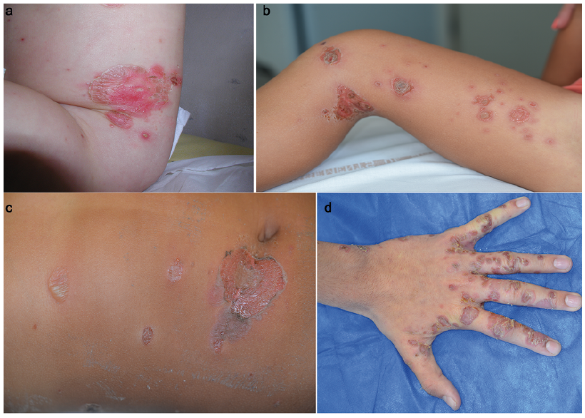
The diagnosis of impetigo is clinical. For S. aureus impetigo, the primary lesion is a fragile bullae. The bullae quickly becomes inflamed and pustular and ruptures to form an oozing erosion or crust (Fig. 1). A frequent and typical localization in children is around the mouth, but any area of the skin can be affected. The grouping of multiple lesions can result in polycyclic erosions with circular contours. The general physical state is preserved. There is no fever; sometimes a satellite lymphadenopathy may be present. Several classical variants have been reported, such as giant impetigo, miliary impetigo, pustules, and dry impetigo. Impetigo neonatorum, previously known as pemphigus neonatorum, is generalized impetigo in neonates. Impetiginization characterizes the secondary infection by S. aureus of a pre-existing dermatosis, usually affecting the epidermis (e.g. eczema, chickenpox, etc.) or secondary to scratching (e.g. pediculosis, scabies, etc.) that results in impetigo or impetigo-like crusted and oozing lesions. A clinical variant of impetigo is ecthyma, in which deep ulceration forms in the dermis (more frequently with S. pyogenes). Scabies is a major cause of impetigo in children worldwide and, more specifically, in disadvantaged populations (6–8). Mass or individual treatment of scabies results in a decrease in the prevalence of impetigo in a community (6–8).
Fig 1. Different clinical presentations of impetigo: a), large dry erosive plaque on adbomen; b) crusted and oozing erosions on the lower limb; c) bullous and oozing erosive lesions on abdomen; d) multiple dry erosions of the hand.
The pathophysiology of staphylococcal impetigo is related to the local production of exfoliatin toxins A and B (1, 9–11). The target protein of exfoliatins A and B is desmoglein 1, a desmosomal protein whose role is the cohesion between keratinocytes, and it is mainly located in the most superficial layer of the epidermis (1, 9–11). The main consequence of the action of the toxin on desmoglein 1 is rupture of keratinocyte cohesion and formation of a bullae. Although bullae are not usually reported in impetigo caused by S. pyogenes, a similar mechanism could be involved; the streptococcal pyrogenic exotoxin B (SpeB) has been demonstrated to be a proteolytic factor that cleaves the extracellular domains of desmoglein 1 and 3 (12).
According to Koning et al. (13) treatment with topical mupirocin and topical fusidic acid are equally effective to, or more effective than, oral treatment, except in extensive impetigo where research is lacking. Penicillin was not as effective as most other antibiotics (12, 13). Hygiene measures, such as strict attention to handwashing, must be applied to prevent recurrence and cross-transmission.
Regarding the risk of antibiotic resistance in impetigo, rare clones of methicillin-resistant S. aureus (MRSA) producing ETA and/or ETB and have been described, mainly from Japan (14–17). In terms of resistance in impetigo, the main concern is with fusidic acid. Resistance to fusidic acid has increased in the early 2000s in some countries of northern Europe, namely Sweden, Norway and the UK. This increase appears to have resulted from the clonal expansion of a strain designated the Epidemic European Fusidic acid resistant Impetigo Clone (EEFIC), which carries the fusidic acid resistance determinant fusB on its chromosome. The high level of use of fusidic acid ointment has been linked to the emergence and spread of fusidic acid resistant S. aureus (18–20).
Folliculitis
S. aureus is responsible for the majority of cases of folliculitis (infection of the pilosebaceous follicle).
Superficial folliculitis. In this condition the infection is restricted to the superficial part of the pilosebaceous follicle (follicular ostium). Clinically it manifests as a pustule, centred on a hair associated with a peri-follicular erythema. All parts of the body with high-density hair can be affected: thighs, perineum, arms, back, eyelid (stye). Sycosis barbae (Fig. 2), whose spread is favoured by shaving, is a particular clinical form localized on the face, characterized by extensive and chronic lesions. Differential diagnoses include folliculitis caused by other microorganisms, such as dermatophytes in kerions, Candida albicans in candida folliculitis, Malassezia folliculitis, Gram-negative folliculitis, non-infectious folliculitis (including Behcet’s disease) and hidradenitis suppurativa.
Fig. 2. Sycosis barbae.
Furuncle (boil). Furuncles, or boils, are characterized by a deep and necrotizing form of folliculitis with involvement of the pilosebaceous follicle in its entirety. It presents as a painful inflammatory papule or nodule, centred around a pustule on a hair-bearing area (the hair has usually disappeared due to necrosis) (Fig. 3). Within a few days of maturation pus will form, associated with necrosis (21). A circular desquamative flange may surround the necrotic centre (22). In recent years it has been found that up to 90% of the S. aureus, isolated from furuncles in some areas produce PVL virulence factor (23–26). This leucocidin leads to local destruction of leucocytes with the formation of larger skin lesions, which respond less well to treatment and tend to recur; the organisms can also cause suppurative pneumonia.
Fig. 3. Furuncle.
The term “furuncle” has sometimes been used in the literature for skin infection caused by other bacteria, such as non-tuberculous mycobacteria (27), but, to avoid confusion, should be reserved for S. aureus infection.
A clinical variant of a furuncle is the carbuncle, defined as a cluster of furuncles. Chronic furunculosis is characterized by the repeated formation of furuncles on different parts of the body over several months (21).
Many reports of systemic infection secondary to a furuncle are reported, but this appears to be rare relative to the high frequency of furuncles. Facial malignant sta-phylococcal infection is a classically described infection, but nowadays it is an exceptionally rare complication of a peri-nasal furuncle leading to a septic facial venous thrombosis that can extend to the cavernous sinus (28).
Abscess
An abscess is a collection of pus. The abscess forms from a tender inflammatory and extremely painful erythematous nodule or plaque. After a few days of evolution, the consistency changes and become soft, indicating the formation of the collection of pus (Fig. 4). Abscesses can be primary or secondary. There is no clearly defined size in the literature for an abscess, therefore in primary abscess, the distinction between a large furuncle and a small abscess is difficult or artificial. Fever is rare, cellulitis, lymphangitis, and satellite adenopathies may be associated. The general physical state is preserved. Pus may appear after some days of spontaneous evolution, and if not drained, spontaneous skin necrosis with rupture and drainage of the pus may occur.
Fig. 4. Primary abscess.
S. aureus is by far the main infectious bacteria isolated from abscesses. The majority of primary or spontaneous abscesses are caused by S. aureus producing PVL (23, 29–31). Secondary abscesses (accidental direct inoculation, drug addiction, septic injections, etc.) are most often, but not exclusively, due to S. aureus (32).
The treatment of suppurative skin infections is based on incision and drainage. The role of antibiotics has been summarized in recent important studies. In the study by Daum et al. (33), 786 participants with a skin abscess 5 cm or less in diameter were treated by incision and drainage and were randomly assigned to receive clindamycin, trimethoprim–sulfamethoxazole (TMP-SMX), or placebo for 10 days; the cure rate among participants in the clindamycin group was similar to that in the TMP-SMX group (221 of 266 participants (83.1%) and 215 of 263 participants (81.7%), respectively; p = 0.73), and the cure rate in each active treatment group was higher than that in the placebo group (177 of 257 participants (68.9%), p < 0.001 for both comparisons). Among the participants who were initially cured, new infections at 1-month follow-up were less common in the clindamycin group. Talan et al. (34) compared TMP-SMX with placebo after incision and drainage of abscesses; clinical cure of the abscess occurred in 507 of 630 participants (80.5%) in the TMP-SMX group vs. 454 of 617 participants (73.6%) in the placebo group (p = 0.005). TMP-SMX was superior to placebo, resulting in lower rates of subsequent surgical drainage procedure, skin infections at new sites, and infections in household members.
Emergence of suppurative skin infection due to community-acquired methicillin-resistant S. aureus. Methicillin has been available since 1961, it was the first semi-synthetic penicillin resistant to penicillinase produced by most of S. aureus at that time (35). Its introduction was quickly followed by the appearance of MRSA (35). This resistance is linked to the synthesis of a modified penicillin-binding protein with less affinity to betalactams, PLP2a, leading to resistance to all beta-lactams (except for new cephalosporins ceftaroline and ceftobiprole). The synthesis of this PLP2a is under the control of the mecA gene, located on a chromosomal mobile genetic element, called the staphylococcal cassette chromosome mec or SCCmec, bordered at both ends by genes called chromosome cassette recombinase (ccRA/ccRB or ccRC), which allow horizontal transmission between and within species. Described almost exclusively in hospitals, these hospital-acquired methicillin-resistant (HA-MRSA), clones have spread widely throughout the world. Over time, they have acquired, in addition to the mecA gene, other resistance genes against other classes of antibiotics, such as macrolides, fluoroquinolones or aminoglycosides (1). However, these clones are rarely involved in skin infections, except for nosocomial operative site infections.
The epidemiology of MRSA has entered a new era the last 25 years. MRSA with new characteristics have emerged in the community setting, namely outside of healthcare facilities (35–39). First reported in Oceania (Australia and New Zealand), these CA-MRSA are currently present worldwide (35–39). Most strains (80–90%) are isolated from suppurative skin infections (35–39). CA-MRSA infections have specific characteristics that clearly distinguish them from HA-MRSA (35–39); they preferentially affect a young population with no previous medical history (35–39). Unlike HA-MRSA, which are often multi-resistant, CA-MRSA generally remains sensitive to most antibiotics apart from beta-lactams. The genetic origin of CA-MRSA is different, with a few major clonal complexes with relative geographical specificity (35–39), USA 300 being the major clone in the USA. The main SCCmec cassettes for HA-MRSA (SCCmec I, II and III) are significantly longer than those for CA-MRAS (mainly SCCmec IV and V). Almost all of CA-MRSA, including the major clones, produce the PVL toxin, which explains the predominance of suppurative skin infections as clinical presentations of CA-MRSA infections. There are no clinical data to suggest that PVL CA-MRSA skin infections differ from PVL methicillin-sensitive ones (MSSA) and their relative prevalence varies in different countries. As CA-MRSA is isolated mainly from suppurative skin infections, the best way to study its epidemiology is to study those infections. Indeed, some countries, such as the USA, have a high rate of CA-MRSA, at approximately 50% of strains isolated (most USA 300) (40–42) and others have a low rate, at less than 10% (43–45). Outbreaks of CA-MRSA are regularly described mainly in different community settings (military personnel, sports teams, drug users, homosexuals, isolated communities, families, etc.) (46–50).
Acute suppurative paronychia
Acute suppurative paronychia is an acute infection of the eponychial nail folds of the hand or foot. Several bacteria may be implicated, but S. aureus is the most common one. The treatment is based on surgical excision; antibiotic treatment plays a minimal role (51).
Lymphangitis
Lymphangitis is caused mainly by S. aureus or S. pyogenes. It is clinically characterized by an erythematous inflammatory linear band, which usually starts from the origin of the infection towards the draining regional lymph node, namely a local adenopathy (Fig. 5). Lym-phangitis is sometimes accompanied by fever. Otherwise, general health state is preserved. Treatment is based on systemic antibiotic therapy.
Fig. 5. Lymphangitis.
Superficial septic thrombophlebitis
An important feature in the pathophysiology of S. aureus infections is its thrombotic capacity. The constitution of a vascular thrombosis allows the infection to develop and cause septic emboli and secondary locations. Staphylococcal skin infection can cause septic thrombophlebitis of the superficial venous network, which can spread to the deep veins. In hospitals, this is most often a complication related to the infection of intravenous catheters. Septic thrombophlebitis is characterized by an inflammatory indurated cord, which begins at the infected site (Fig. 6). Treatment is based on antibiotic therapy and treatment of the portal of entry. A particular form of such thrombophlebitis is facial malignant staphylococcal infection (see above).
Fig. 6. Thrombophlebitis from catheter site.
Cellulitis
Cellulitis may occur associated with an abscess or a thrombo-phlebitis or complicate an acute or chronic wound as a result of secondary infection. It is more common with S. pyogenes. The treatment is based on systemic antibiotic therapy.
Necrotizing fasciitis
A few reports of necrotizing fasciitis (NF) associated with S. aureus have been published. Miller et al. (52) reported 14 cases in 2005 caused by CA-MRSA. A few other isolated cases have been published since. Given the scarcity of the reports, NF caused by S. aureus seems exceptional.
Contiguous infections
These are related to a suppurative focus located near the skin (53). They manifest as an inflammatory mass that simulates an abscess, particularly in the vicinity of septic arthritis, osteomyelitis, bursitis, tenosynovitis or infected false aneurysms or myositis. Sometimes cutaneous fistulization occurs.
Secondary infections of acute or chronic wounds
They are a common situation in practice. Clinically, secondary infections show local inflammatory signs (pain, erythema) or cellulitis, and the possible presence of pus (54). The isolation of S. aureus in a wound is not synonymous with local infection, but must be interpreted according to the clinical presentation and, especially, the presence of inflammatory signs. The distinction between secondary infection and colonization may be difficult.
Botryomycosis
S. aureus can cause botryomycosis, a rare, chronic and granulomatous infection characterized by painless slow-growing papulonodules, abscesses and ulcers and, histopathologically, the presence of granules composed of bacterial cocci (55).
Toxic shock syndrome
The toxic shock syndrome (TSS) was first described by Todd (1978) in 7 children who had a generalized erythema, fever, hypotension, diarrhoea and multi-organ failure (56). In 1980 many cases were reported in young women who used certain types of tampon (57, 58). The incidence of menstrual TSS in the US peaked in 1980 and has decreased significantly since the removal of these tampons from the market (59).
TSS is due to the production of a toxin by S. aureus, mainly TSST-1 and staphylococcal enterotoxins, particularly enterotoxin B and, less commonly, other enterotoxins (56). The 1997 CDC definition (60) includes the following clinical criteria: fever (≥ 38.9°C) a diffuse macular erythroderma, desquamation (1–2 weeks after onset of illness, particularly on the palms and soles), hypotension, multisystem organ involvement (57). In a study of 130 TSS, Reingold et al. (57) found a skin infection in 30% of cases, a genital focus in 27% (after delivery or abortion), 18% post-surgery focus, and in 13% the source was not identified. The pathogenesis of TSS is linked to the properties of superantigens in S. aureus toxins, namely activation of greater numbers of T lymphocytes resulting in the production of high levels of cytokines (33). Skin manifestations of TSS include a generalized erythema (with palm and sole involvement) (Fig. 7). Palmar, sole and finger desquamation may occur after recovery (Fig. 8). Transient alopecia, nail shedding and increased sweating on the hands and feet have been described (61). Treatment is based on the treatment of the multi-organ failure and the S. aureus focus of infection. Some antibiotics acting as protein-synthesis inhibitors with anti-toxaemic properties could provide additional therapeutic benefits (62).
Fig. 7. Erythema of toxic shock syndrome.
Fig. 8. Distal desquamation after toxic shock syndrome.
In Japan, Takahashi et al. (63) have reported neonates who developed generalized erythema and thrombocytopaenia in the first week of life associated with MRSA-producing TSST-1. They propose neonatal toxic-shock-syndrome-like exanthematous disease (NTED) as the name for this disease. Similar cases have been reported in Europe (64).
“Staphylococcal scarlet fever”
Staphylococcal scarlet fever, also called scarlatiniform erythroderma/rash, was first described in the 1920s. Lina et al. (65) found that 16 out of 17 strains of S. aureus isolated from patients with staphylococcal scarlet fever produced TSST-1, enterotoxins, or both. Enterotoxin B was the predominant toxin involved in a study in Taiwan (66). It is possible that most cases of staphylococcal scarlet fever are, in fact, a mild or attenuated clinical manifestation of TSS.
Staphylococcal scalded skin syndrome
When the ETs spread systemically, they can cause SSSS (9–11, 67). It is a generalized blistering disease affecting mainly neonates and young children and, exceptionally, adults with underlying diseases. The disease begins abruptly with fever and generalized erythema, followed by large fragile blisters involving the entire skin surface within the next few hours to days, which rupture rapidly (with a positive Nikolsky sign) (67). Widespread involvement of the entire skin surface can occur, but the mucous membranes are usually spared. Mild forms of SSSS have been described where the SSSS is limited to the body folds associated with a fine generalized desquamation (68). The disease follows localized S. aureus infection. Poor renal clearance of the toxins by neonates and by adults with impaired renal function is a major risk factor for developing SSSS. The prognosis of SSSS in children, who are appropriately treated, is good, with a mortality of less than 5%, but it may be fatal in up to 60% of affected adults, usually due to underlying diseases (67). The diagnosis of SSSS is clinically based. Exfoliatins are produced by S. aureus at a distant site; the blister fluid in generalized SSSS is usually sterile. The treatment is based on dressings, where there are large blisters, and the eradication of the source of S. aureus infection focus.
Skin manifestations of S. aureus endocarditis
Endocarditis caused by S. aureus is classified as acute endocarditis. The description of endocarditis-related skin manifestations is confusing; Janeway lesions and Osler’s nodes were described at the beginning of the 20th century (1). Classically reported Janeway lesions are macular, purpuric lesions that occur on hands and feet (Fig. 9). Histologically, they show neutrophilic microabscesses in the dermis and vessel thrombosis (69). These lesions are thought to be caused by septic microemboli; results of culture of skin specimens are frequently positive (70–73). Osler’s nodes are described as small, painful, nodular lesions on the fingers or toes. Only a few biopsied Osler’s nodes gave positive results on culture, and histological examination showed diverse findings (73). The description of Janeway lesions corresponds better with the skin manifestations of S. aureus endocarditis.
Skin manifestations of S. aureus bacteraemia (without endocarditis)
Such manifestations related to the frequency of S. aureus bacteraemia are extremely rare. Purpuric disseminated eruptions and abscesses are the main clinical manifestations that have been described as a secondary focus of S. aureus bacteraemia. Exceptionally, purpura fulminans has also been reported (74).
Fig. 9. Purpura during endocarditis.
Immunological manifestations associated with acute or chronic S. aureus infections are rare. A few cases of vasculitis or Henoch-Schönlein purpura have been reported, mainly in the course of S. aureus bacteraemia (75–77). Some of these associations may be coincidental.
Staphylococcal skin infections are part of a complex group of diseases. Unfortunately, most reports in the literature classify skin infections and S. aureus skin infections under the heading “skin and skin structures infections (SSTI)”, giving the illusion that all skin manifestations are within the same clinical spectrum. This review shows, on the contrary, how the clinical spectrum of skin manifestations due to S. aureus is diverse and related to different physiopathologies. Further reports and studies on skin S. aureus infections should take into consideration this rich and varied clinical spectrum of disease.
This review has focused on the clinical and therapeutic aspects of S. aureus skin infections, and many other questions are not mentioned, such as the interactions of S. aureus with the skin microbiome, the reservoirs of S. aureus, the relationships between reservoirs and skin infections, and the decolonization of the reservoirs. All of these complex topics are currently the subject of intense research.


 Click to show fullsize
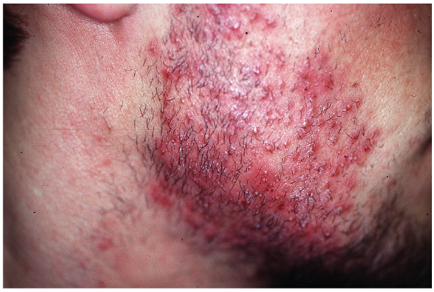 Click to show fullsize
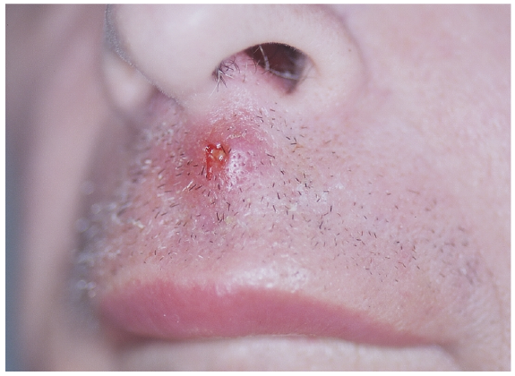 Click to show fullsize
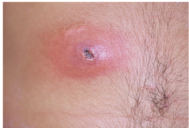 Click to show fullsize
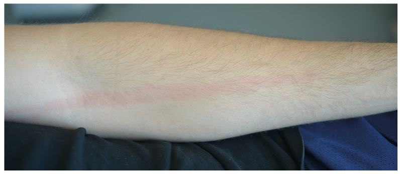 Click to show fullsize
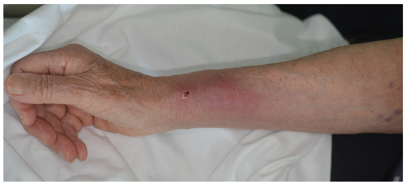 Click to show fullsize
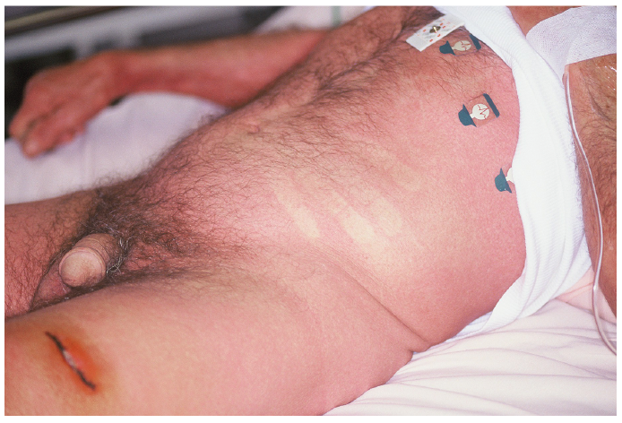 Click to show fullsize
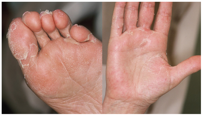 Click to show fullsize
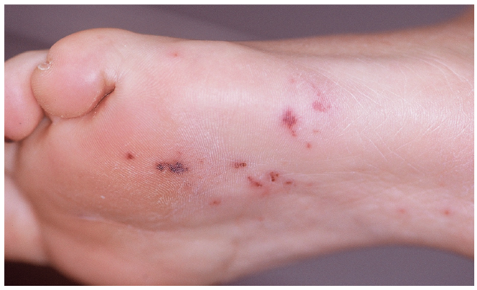 Click to show fullsize