


Department of Dermatology, Venereology, and Allergology, University Medical Center, Ruprecht-Karls-University Heidelberg, Im Neuenheimer Feld 440, DE-69120 Heidelberg, Germany. E-mail: julia.winkler@med.uni-heidelberg.de
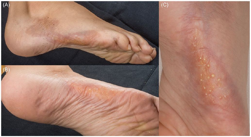
A 9-year-old girl presented to our outpatient clinic with a skin lesion on her right lateral foot (Fig. 1). Her medical history showed that the lesion had been reported since the age of 5 years. The medical history was otherwise unremarkable. The lesion appeared as orange verrucous plaques with keratotic pits and a peripheral erythema distributed along Blaschko’s lines. It extended from the lateral malleolus to digits 4 and 5, which appeared slightly smaller than the toes on the left foot. The patient also reported some restriction in mobility in the corresponding toes. The skin lesion was also sensitive to pressure and touch.
A punch biopsy was obtained and histopathology revealed hyperkeratosis and epidermal acanthosis with prominent cornoid lamella (haematoxylin-eosin (HE) staining ×50, Fig. 2A). Close-up of cornoid lamella revealed accompanying epidermal thinning and reduced stratum granulosum (HE ×100, Fig. 2B).
For diagnostic evaluation of potential orthopaedic abnormalities, magnetic resonance imaging (MRI) was recommended.
In the reported case, numerous topical treatment modalities, including urea, steroids, vitamin D analogues and retinoids, were recommended and evaluated, but with limited success.
What is your diagnosis? See next page for answer.
Fig. 1. Keratotic naevus on the right foot. (A) Lateral view, (B) plantar view, (C) close-up view.
Fig. 2. Haematoxylin eosin (HE) staining shows hyperkeratosis and epidermal acanthosis with prominent cornoid lamella (A: ×50). Close-up view of cornoid lamella accompanied by epidermal thinning and reduced stratum granulosum (B: ×100).
Acta Derm Venereol 2020; 100: adv00201.
Diagnosis: Porokeratotic eccrine ostial and dermal duct naevus (PEODDN)
Porokeratotic eccrine ostial and dermal duct naevus (PEODDN) is a naevoid skin lesion with histopathology revealing cornoid lamella, which is therefore classified as a porokeratotic dermatosis. This uncommon eccrine hamatoma is localized mainly on the distal extremities presenting with verrucous lesions distributed along Blaschko’s lines (1). It generally appears in childhood and, due to its rare occurrence, may pose diagnostic difficulties. However, the histopathology is quite characteristic, showing cornoid lamellae, which define porokeratosis (2). The current case presents typical clinical and histopathological characteristics of this rare entity. Case reports in English language are limited (2). Thus, in clinical routine PEODDN may easily be misdiagnosed. The diagnosis may only be made after clinicopathological correlation. Differential diagnoses include epidermal naevi, such as linear and verrucous epidermal naevus, linear forms of psoriasis, naevus comedonicus, punctate palmoplantar keratoderma and linear porokeratosis (1). In 1979, PEODDN was first reported by Marsden et al. as comedo naevus of the palm (1). The nomenclature is still discussed; recently whether PEODDN and porokeratotic and eccrine hair follicle naevus may more precisely be summarized as porokeratotic adnexal ostial naevus (3). With regard to aetiology of PEODDN a genetic mosaicism is discussed due to expansion along Blaschko’s lines. Mutations in the gap junction protein connexin 26 have been reported (4). In lesional epidermis homozygous mutation was reported (4). Since connexin 26 mutations are found in keratitis-ichthyosis-deafness syndrome, PEODDN may represent a related cutaneous mosaic variant (4). For our patient genetic counselling was initiated and a genetic mosaic considered likely, but to date no further biopsy has been performed. Medical conditions reported in association with PEODDN include hyperthyroidism, polyneuropathy, deafness, developmental delay, seizure, scoliosis and squamous cell carcinoma (1). Clinical appearance concerning expansion and extent of keratinization may vary (5). The current case is typical concerning onset in childhood and unilateral plantar localization (2). Even more characteristic than clinical appearance are histopathological features. Cornoid lamellae, which define cutaneous porokeratosis, in association with eccrine acrosyringia, may be considered diagnostic for PEODDN (2). Treatment options discussed include topical steroids, calcipotriol, phototherapy and cryotherapy. Moreover, lesions may be surgically removed or treated with CO2 or erbium laser (5). Nevertheless, treatment results often remain unsatisfactory, and spontaneous involution is rare.


 Click to show fullsize
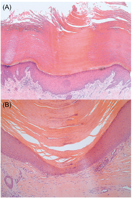 Click to show fullsize