


1Department of Dermatology and 3Laboratory of Pathology, Assistance Publique – Hôpitaux de Paris, Hôpital Avicenne, 2University of Paris XIII, Sorbonne Paris Cité, Bobigny, 4Laboratory of Pathology and 5Department of Dermatology and INSERM U1058, University of Montpellier Montpellier France. E-mail: o-dereure@chu-montpellier.fr
Accepted Jun 1, 2020; Epub ahead of print Jun 9, 2020
Acta Derm Venereol 2020; 100: adv00226.
Several studies have suggested a higher risk of occurrence of lymphoproliferative disorders (LPDs) in patients with rheumatoid arthritis (RA) treated with MTX, a subset of conditions overall known as MTX-associated LPDs and categorized by the WHO as “other iatrogenic immunodeficiency-associated LPDs” (OIIA-LPD) (1–3). Among them, diffuse large B cell lymphoma (DLBCL), either related to Epstein Barr virus (EBV) or not otherwise specified (NOS) and classical Hodgkin disease are the main subtypes, followed by polymorphic/lymphoplasmacytic infiltrates (P/L-I), Hodgkin-like lesions (HLL) and EBV+ mucocutaneous ulcer. MTX-associated LPDs initially presenting with skin lesions have been reported in a minority of cases, and mainly identified as primary cutaneous B-cell lymphoma in single case-reports or small series (1, 4, 5) with only 4 cases in patients with prior CTCL (6–9). We report here on 3 further cases of primary cutaneous B-cell LPD occurring in a setting of CTCL treated with MTX.
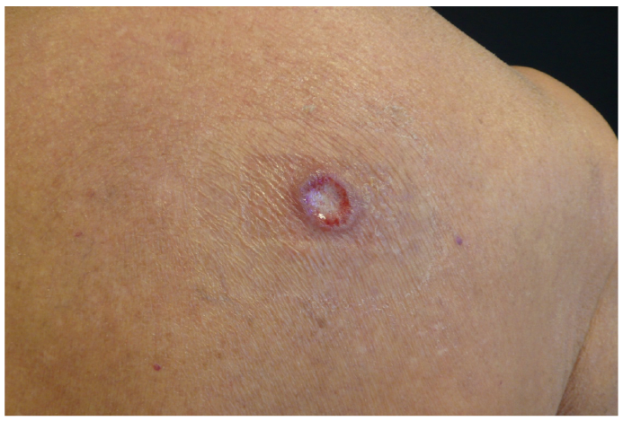
Patient 1. An 80-year-old woman was diagnosed with erythrodermic mycosis fungoides (MF) stage IIIA (T4N1M0B0b). Ten months after MTX introduction as first-line treatment with an initial weekly dosage of 20 mg rapidly increased to 40 mg with a favourable response, she presented with a solitary ulcerated nodule of the upper back (Fig. 1) with no further LN enlargement or hepatosplenomegaly. Absolute total lymphocytes cells counts was 0.860 G/l. Skin biopsy of the nodule displayed scattered CD3+ CD4+ CD5+ CD7– CD8– T-cells documenting residual MF admixed with a heavy dermal infiltrate consisting of large atypical CD20+ Pax5+ lymphocytes with strong EBER expression on in situ hybridization (ISH) (Fig. S1) and B clonality. EBV viral load was also detected in peripheral blood (3.5 log). The diagnosis of EBV+ primary cutaneous diffuse large B-cell lymphoma (PCDLBCL) associated with MTX treatment of the underlying CTCL was considered, and MTX withdrawal resulted in a total disappearance of the skin nodule within one month. The patient remained relapse-free after a 6-year follow-up, but eventually died from a non-CTCL-related cause.
Fig. 1. Clinical presentation of patient 1. Solitary ulcerated nodule on the upper back 10 months after introduction of methotrexate.
Patient 2. A 78-year-old man with Sézary syndrome (SS) developed twenty months after MTX implementation at a weekly dosage of 15 mg increased to 22.5 mg a 3-cm right temporal ulcerated nodule quickly developed, followed by occurrence of 4 similar but smaller, ulcerated nodules on the scalp with no general status alteration or LN changes. Absolute total lymphocytes and Sézary cells counts were 1.150 G/l and 0.770 G/l, respectively. Chest, abdomen and pelvis CT scan were unremarkable apart from a few moderately enlarged stable peripheral LN. Biopsy of a scalp nodule revealed a diffuse, dense and monotonous dermal infiltrate of large CD20+ Pax5+ B-cells with strong EBER expression on ISH. A dominant B clone was present in skin nodules, but not in peripheral blood. Bone marrow biopsy was normal. No Myd88 mutation was identified. A diagnosis of EBV+ PCDLBCL associated with MTX was retained. Replacement of MTX by bexarotene, topical superpotent dermocorticoids and extracorporeal photochemotherapy ECP resulted in a total disappearance of skin nodules in 3 months with no relapse of the B cell lymphoma after an 8-year follow-up.
Patient 3. A 64-year-old woman treated for SS with MTX alone (15 then 22.5 mg weekly) subsequently associated with bexarotene and ECP suddenly developed after 5 years of this therapeutic regimen a relapsing pruritic erythroderma, massive infiltration of the right arm, multiple ulcerated nodules of the chest and the right axilla and multiple enlarged lymph nodes. No visceral involvement or deep LN enlargement was identified by imaging procedures, but haematological parameters worsened with 2.750 G/l of circulating Sézary cells for an absolute total lymphocyte count of 3.400 G/L and a CD4/CD8 ratio of 70. Multiple skin biopsies obtained from chest nodules and right arm revealed a diffuse, dense dermal infiltrate of small- and medium-size atypical CD3+ CD4+ CD5+ CD7– CD8– T cells admixed with large CD20+ Pax5+ atypical B cells. No EBV expression identified by both IHC and ISH. An identical dominant T clone was present in skin and peripheral blood, and a B clone was identified in skin nodular lesions, but absent in peripheral blood. LN biopsy of the axillary mass displayed the same T cell proliferation as in skin lesions, but not the B-cell proliferation identified in skin nodules. A diagnosis of MTX-induced PCDLBCL with no EBV reactivation in association with a severe relapse of SS was primarily considered. MTX was withdrawn and 6 courses of systemic chemotherapy with rituximab, cyclophosphamide, doxorubicin, vincristine and prednisolone (R-CHOP) resulted in total clinical remission of chest nodules and partial remission of SS. Further CTCL progression occurred after a few months with no evidence of PCDLBCL recurrence and the patient eventually died from septic complications 17 months after diagnosis of B-cell lymphoma.
We report 3 new cases of B-cell LPD occurring in patients with CTCL treated with MTX, all of them categorized as PCDLBCL either EBV+ or not with no evidence of systemic disease. In the literature, MTX-associated LPDs are predominantly of the B-cell subset (10) and clinically present as nodal systemic lymphoma with a histologically pattern of DLBCL and strong EBV expression in most cases (4, 5). In these patients, iatrogenic LPD occurrence is therefore presumed to be closely related to EBV reactivation secondary to MTX immunosuppressive effect (11). Treatment of systemic MTX-associated LPDs is primarily based on drug withdrawal, resulting in a favourable course in most cases (12, 13). Conversely, a combined rituximab and polychemotherapy (R-CHOP) regimen may be warranted in some cases owing to a more protracted outcome (10).
Four similar cases of PCDLBCL occurring in MTX-treated CTCL have been previously reported in the literature (6–9). All 7 cases, including ours, were erythrodermic CTCL (4 SS and 3 erythrodermic MF; 5 males/2 females; median age 74 years) and the cutaneous B-cell LPD presented with rapidly evolving cutaneous papules and nodules occurring 2–66 months after MTX introduction (mean 22.8 months) with a close histological pattern of monotonous dermal infiltrate of diffuse large B-cells admixed with features reminiscent of the underlying CTCL in 3/7 cases and positive EBV markers in 6/7 cases (86%). Similarly to MTX-associated systemic LPDs, cutaneous lesions totally vanished in all cases in less than 3 months after MTX discontinuation alone (4/7 patients) or associated with a systemic treatment targeting malignant B cells (3/7 patients: R-CHOP in 2/7 and rituximab alone in 1/7). No relapse was observed after a mean follow-up of 35.8 months (range 3–96 months), but 3/7 patients eventually died from CTCL evolution. All patients’ characteristics and outcome are summarized in Table SI. An additional role of lymphoma-related immunosuppression in LPD pathomechanisms cannot be ruled out especially in SS, as illustrated by our EBV negative case.
Lymphocytes have been suggested to play a key role in the pathogenesis of MTX-associated LPD, with a decrease in the absolute lymphocyte count in peripheral blood at the time of LPD development (14). Interestingly, the total count of normal lymphocytes was low in all our patients at the time of PCDLBCL diagnosis and increased rapidly after MTX discontinuation in patient 2 concomitantly with definitive regression of cutaneous lesions.
In conclusion, the onset of skin tumour(s) or nodule(s) in patients with CTCL treated with MTX should alert to the possibility of MTX-induced primary cutaneous B-cell lymphoma. Treatment is primarily based on drug withdrawal, resulting in a favourable course in most cases.


 Click to show fullsize