


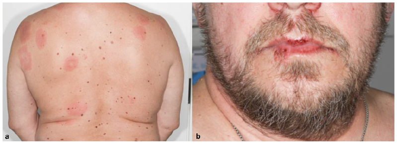
A 56-year-old man with hypertension and marginal zone B-cell lymphoma (which had been treated with cytostatic drugs in 2014 and 2015 and had subsequently been in remission for 3 years) was referred in 2019 to the Department of Dermatology, Oulu University Hospital, presenting with multiple red lesions on the skin. The patient had been bitten on the right hand by his dog 1 week earlier. Skin lesions appeared 6 days after the dog bite. The patient had fever, but he was otherwise well. In laboratory tests the level of C-reactive protein was 46 mg/l and leucocytes 8.7 × 10⁹/l. Physical examination revealed sharply demarcated red lesions 2–5 cm in diameter on his back (Fig. 1a), thighs and scalp. The lesions were warm to the touch and some were clear in the centre. The bite had left only a minor mark on the patient’s right hand. He had manifestation of herpes simplex infection on his lips (Fig. 1b). He had received acyclovir prophylaxis during his cytostatic treatment, which had been discontinued. There were no symptoms of mucous membrane involvement. No enlarged lymph nodes were found. A punch biopsy of a skin lesion revealed perivasculitis, lympho-plasmacytic inflammation and some eosinophils.
What is your diagnosis? See next page for answer.
Fig. 1. (a) Several sharply demarcated red lesions on the back. (b) Erosions and crusting typical for herpes simplex on the lips.
Acta Derm Venereol 2020; 100: adv00218.
Diagnosis: Erysipeloid, diffuse cutaneous form and systemic infection
Erysipeloid is an acute bacterial infection of the skin and other organs caused by the micro-organism Erysipelothrix rhusiopathiae. E. rhusiopathiae can cause self-limited soft tissue infection or serious systemic infections. This Gram-positive, rod-shaped bacterium is widespread in nature and is found in many wild and domestic mammals, birds and fish. The bacterium is also found in contaminated water, soil and food products. Erysipeloid in humans often takes the form of an occupational dermatosis acquired after direct contact between injured skin and infected animals or material. Farmers, people working in the fishing industry, veterinarians and slaughterhouse workers have the highest risk of infection (1, 2).
Cutaneous E. rhusiopathiae infection has 3 major clinical manifestations in humans. Localized cutaneous infection (erysipeloid) is the most common and the least severe form. Skin lesions appear first at the trauma site and develop slowly over a few days from small red macules to more violaceous lesions. These typically appear on the fingers, dorsal hands or forearms. The inoculation time is 1–7 days. The macules are clearly defined, with raised borders, and often display central healing. Patients may feel pain or burning in the lesions. Swelling of affected fingers is representative, while systemic symptoms are uncommon. Diffuse cutaneous infection is rarer. It is characterized by multiple skin lesions, which can be urticarial or bullous. Fever, malaise, headache and arthralgia are common, but blood cultures are usually negative. This form is seen more often in immunocompromised patients. Both the localized and diffuse cutaneous forms are self-limiting and resolve spontaneously within one month. However, treatment with antibiotics is recommended to enhance healing and to prevent the disease from spreading to other organs (1–3).
Systemic infection means that the infection has spread to organs other than the skin. The most common sites of manifestation of a systemic infection are the heart, brain, joints and lungs. Endocarditis is seen in 90% of patients with a systemic E. rhusiopathiae infection. Patients often present with fever, and blood culture is positive for E. rhusiopathiae. Skin lesions are generally found on the trunk and extremities, but may also be absent. Chronic liver disease and immunosuppression increase the risk of systemic disease (1–3).
Diagnosis of E. rhusiopathiae infection is based on typical clinical findings alongside a history of exposure to infected animals or material. E. rhusiopathiae can be isolated from the blood with a routine blood culture. Histopathological findings are non-specific; oedema in the dermis, vascular dilatation, and an inflammatory infiltrate consisting mainly of neutrophils and lymphocytes, with some eosinophils are often seen. However, skin biopsy is rarely needed because of the classical clinical picture (4).
In case described here, a more detailed anamnesis revealed that the patient had been bitten while trying to remove the rotten carcass of a bird from his dog’s mouth. One week later he started to experience fever, and skin lesions appeared. His blood culture was positive for E. rhusiopathiae. The patient was hospitalized. The drug of choice in erysipeloid infection is penicillin, but because of the suspicion of a penicillin allergy our patient was treated with intravenous clindamycin and responded well. Topical treatment (betamethasonepropionate cream) was used for the skin lesions. The patient’s cardiac ultrasound was normal and there were no clinical symptoms of endocarditis. It is possible that his history of mucosa-associated lymphoid tissue lymphoma and its treatment with cytostatic drugs predisposed our patient to the disseminated and systemic form of erysipeloid. Fortunately, his disease did not manifest in any organs other than the skin.


 Click to show fullsize