


1Department of Dermatology, 2Western Finland Cancer Centre (FICAN West) Cancer Research Laboratory and 3Auria Biobank, University of Turku and Turku University Hospital, Turku, Finland
Cutaneous squamous cell carcinoma (cSCC) has metastatic potential. The aims of this study were to identify the risk factors for metastasis of primary cSCC and for poor prognosis in metastatic cSCC. Retrospective primary tumour cohorts of metastatic cSCCs (n = 85) and non-metastatic cSCCs (n = 218) were analysed. The mean annual rate of metastasis for primary cSCCs was 2.28%. In 49.4% of patients with metastatic cSCC, metastasis was detected within 6 months of diagnosis of the primary cSCC. There was no prior history of cSCC in 84.7% of metastatic cSCCs. Risk factors for metastasis included Clark’s level 5, tumour diameter 20–29.9 mm, age at diagnosis < 50 or 70–79 years, and location on lower lip or forehead. A reduced risk of metastasis correlated with: isosorbide mono-/di-nitrate and/or aspirin use; comorbidity with actinic keratosis or basal cell carcinoma; and actinic keratosis or cSCC in situ as part of, or confirmedly preceding, primary cSCC. Poor prognosis in metastatic cSCC correlated significantly with ≥ 3 nodal metastases and extranodal extension of metastasis. These results characterise new risk factors for metastatic cSCC.
Key words: keratinocyte carcinoma; metastasis; actinic keratosis; cutaneous squamous cell carcinoma; basal cell carcinoma; skin; cancer.
Accepted Sep 7, 2020; Epub ahead of print Sep 14, 2020
Acta Derm Venereol 2020; 100: adv00266.
doi: 10.2340/00015555-3628
Corr: Veli-Matti Kähäri, Department of Dermatology, University of Turku and Turku University Hospital, Hämeentie 11 TE6, FI-20520 Turku, Finland. E-mail: veli-matti.kahari@utu.fi
The incidence of cutaneous squamous cell carcinoma is increasing worldwide. Cutaneous squamous cell carcinoma has metastatic potential and causes mortality. However, clinical assessment of the risk of metastasis is challenging, and the risk factors have not been established. This study shows that metastasis of primary cutaneous squamous cell carcinoma occurs early, and that there is no prior history of cutaneous squamous cell carcinoma in the majority of cases. The presence of previous cutaneous squamous cell carcinoma precursors and basal cell carcinoma reduces the risk of metastasis. This study provides novel evidence that the use of aspirin and/or isosorbide mono-/di-nitrate correlates significantly with a lower risk of metastasis. These findings provide new risk and prognostic factors for metastatic cutaneous squamous cell carcinoma at the patient and tumour level.
Cutaneous squamous cell carcinoma (cSCC) is a keratinocyte carcinoma with increasing incidence worldwide. It is the most common skin cancer with metastatic potential (1, 2). In Finland the incidence of cSCC has doubled over the past 10 years (3). The overall rate of metastasis has been estimated as 1–4%, and cSCC accounts for at least 20% of all skin cancer-related mortality worldwide (2, 4–6). The prognosis for patients with metastatic disease is generally poor, and mortality correlates primarily with nodal metastases (7, 8).
Cumulative solar UV radiation is the main aetiological factor for cSCC. cSCC lesions typically develop on sun-exposed areas of skin, most frequently in the head and neck region of fair-skinned elderly individuals (9). The presence of UV radiation-induced precursor lesions, actinic keratoses (AKs), in previously unaffected individuals has been reported as one of the strongest predictors for the development of cSCC (10).
Established tumour (T) staging systems, published by the American Joint Committee on Cancer (AJCC) and the Brigham and Women’s Hospital (BWH), are used in clinical risk stratification. Tumour diameter, invasion depth, applied as invasion beyond fat or Breslow thickness, and perineural invasion, correlate with the risk of metastasis and are used as classifying factors in both the BWH and the 8th edition of AJCC (AJCC-8) staging systems (11). However, it has been suggested that current staging systems are unsatisfactory in predicting the progression of primary cSCC to metastatic disease (12). Other proposed risk factors in correlation with metastasis include lymphovascular invasion, certain histological subtypes, differentiation grade, local recurrence, increasing number of cSCCs, immunosuppression, and certain locations and location properties (11, 13–15). However, there is controversy regarding the value of these factors in predicting the risk of metastasis of cSCC, as these findings are based mainly on retrospective single-institution studies with small cohorts and there is wide variation in study design and the reporting of results.
The aims of this study were to identify factors that correlate with the risk of metastasis of primary cSCC and with poor prognosis of metastatic cSCC (mcSCC) at the patient and tumour level by generating and analysing cohorts of mcSCC and non-mcSCC. To date, there are few studies comparing larger cohorts of mcSCCs in comparison with non-mcSCCs, and none with as comprehensive analysis of risk factors at the patient level (2, 12, 16–18). The results of the current study characterise new risk factors for metastatic cSCC.
Ethical approval
The study was approved by the Ethics Committee of the Hospital District of Southwest Finland (study number 4/2006). Registry study approval for the collection and use of clinicopathological data were provided by Scientific Steering Committee of Auria Biobank (study number AB15-9721) and Turku University Hospital Clinical Research Centre (study number T80/2018). The study was performed in accordance with the principles of the Declaration of Helsinki (19). Finnish Cancer Registry data (3) is in the public domain, and was utilized according to national legislation.
Research material
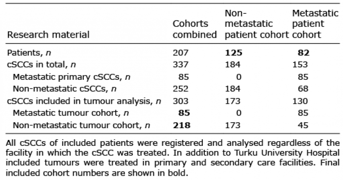
The area served by Turku University Hospital constitutes the study region. All patients in the region who require tertiary care centre-level treatment for mcSCC are treated at Turku University Hospital. To identify all patients with mcSCC in the study region, 3 sequential automated screenings were carried out by Auria Biobank, using the topographical code C44 (ICD-10) and keywords “squamous” and “metastatic”. The screening identified 992 patients, of whom 856 had potential mcSCC (Fig. S1). A manual review of the patient records and pathology reports resulted in a final total of 207 patients being included in the study. These 207 patients were divided into 2 cohorts: a metastatic cohort of 82 patients with at least one mcSCC and a non-metastatic cohort of 125 patients with solely non-mcSCC with no metastasis during a 5-year follow-up (Table I). From the above patients with 337 primary cSCCs, 324 tumours were eligible for tumour analysis, based on the availability of pathology reports. Furthermore, non-mcSCCs diagnosed within less than 5 years of data collection were excluded. Four patients in the non-metastatic patient cohort had cSCC classified as locally advanced and unresectable, one of whom was excluded from tumour-level analysis due to the ambiguous nature of the case. As a result, 303 tumours (85 individual mcSCCs and 218 non-mcSCCs) from 206 patients constituted the tumour cohorts (Table I).
Table I. Summary of research material. Non-metastatic cutaneous squamous cell carcinoma (cSCC) patient cohort (n = 125), metastatic patient cohort (n = 82); non-metastatic tumour cohort (n = 218), metastatic tumour cohort (n = 85)
Patient and tumour variables
Clinical and histopathological data were gathered manually from the patient records and pathology reports between 1 June 2018 and 22 August 2019. At the patient-level age, sex, medication and comorbidities, as well as information on smoking, occupation, diagnostics, treatment and survival were collected. At the tumour level, age at the exact time of tumour diagnosis, sex, histopathological and clinical tumour characteristics for both the primary tumours and metastases containing every biopsy, excision and re-excision were collected.
Inclusion criteria regarding medication for patients with non-metastatic disease was long-term (at least 6 months’) regular use of medication prior to diagnosis of the first cSCC and, for patients with metastatic disease, prior to diagnosis of the first primary mcSCC. Comorbidities were registered if diagnosed prior to diagnosis of the first cSCC or first mcSCC, respectively. Co-malignancies and keratinocyte premalignancies were included whether diagnosed prior to or after the diagnosis of first cSCC or mcSCC. Solid-organ transplant recipients (SOTRs), patients on immunosuppressive medication, those with HIV infection, chronic lymphocytic leukaemia, or non-Hodgkin’s lymphoma were considered immunocompromised.
In survival analysis death was the primary clinical endpoint. The exact cause of death was rarely reported and autopsies were infrequently performed. Therefore, unambiguous overall survival (OS) was chosen for analysis of survival. Death or last contact with healthcare provider represented the end of follow-up.
The date of the first cSCC tissue specimen was selected as the date of diagnosis of primary tumour. Primary tumours in the tumour cohorts were diagnosed during the period 1993 to 2018. Local recurrences were regarded as a continuum of the primary tumour and not counted as individual cSCCs. The date of diagnosis of metastasis was either the date of imaging study or the date of tissue specimen, depending on which was performed first. Concerning metastases, all mcSCCs were histopathologically confirmed, except for 2 primary mcSCCs, which had solely radiologically diag-nosed metastases. For tumour data analysis of non-mcSCCs, the presence of characteristics at any point of the tumour continuum was sufficient for inclusion. For primary mcSCCs, characteristics present prior to, or at the time of, detection of metastasis, were included in tumour analyses. For non-mcSCCs the highest values of variables observed at any point of the tumour continuum were chosen. For primary mcSCCs the highest values of variables observed prior to, or at the time of, detection of metastasis were chosen. Quantitative invasion depth was infrequently (25.7%) expressed in pathology reports, but Clark’s level could be deduced from the pathology reports for 96.0% of tumours with a level of accuracy segregating Clark 2–4 from Clark 5.
For treatment of mcSCCs, complete response was achieved if both the primary tumour and all metastases were treated successfully and no disease activity occurred during follow-up.
Parotid gland metastases were interpreted solely as nodal in origin. Extranodal extension (ENE) was registered when mentioned in the clinical record or pathology report.
Both AJCC-8 and BWH primary tumour staging were determined utilizing characteristics present prior to, or at the time of, metastasis detection. AJCC-8 was applied only to tumours located in the head and neck region.
Rate of metastasis
The Finnish Cancer Registry registers and receives information on every cancer detected and treated in Finland. This public database was used to determine the metastatic rate of cSCC in the study region (3). The number of mcSCCs was distributed based on the year of the corresponding primary mcSCC diagnosis, divided by the incidence of cSCC in the study region for each year. In the determination of metastatic rate the study included cSCCs that were diagnosed during the 10-year period 2004 to 2013, thus including cSCCs with more than 5 years for development of metastasis.
Statistical analysis
All statistical analyses were conducted using IBM SPSS Statistics for Windows, version 25.0 (IBM Corp., Armonk, NY, USA). Bidirectional p-values < 0.05 and 95% confidence intervals (CIs) of odds ratios (ORs) not including 1.00 were considered statistically significant. For patient-level analyses, all patients were counted once, regardless of the number of individual cSCCs. For tumour-
level analyses every cSCC was counted once regardless of the patient cohort, number of tissue specimens, or local recurrences. Baseline patient and tumour characteristics were analysed using descriptive statistics mainly crosstabs and frequency tabulation. Statistical analyses were conducted with Pearson χ2 test and Fisher’s exact test. For scale variables, such as age, the Mann–Whitney U test or independent sample t-test was applied, with interquartile ranges (IQR) and standard deviations (SD), respectively.
Binary logistic regression analyses with 95% CIs were performed in order to determine ORs regarding the risk of metastasis and prognosis of mcSCC. Variables were selected for logistic regression analyses if descriptive statistical analyses showed statistical significance (Tables SI, SII) or if it was reasonable based on clinical parameters (Patient level: age, sex, immune status, SOTR, cSCCIS; Tumour level: sex). For tumour-level analyses, logistic regressions were performed using generalized estimating equations, in order to estimate the impact of multiple tumours on the same patient. Adjusted ORs (aORs) were calculated, including variables with significant crude ORs. Staging systems were excluded from adjusted models due to clinical reasoning and multicollinearity.
The Kaplan–Meier method was used to generate survival curves and define survival probabilities (20). Statistical analysis was performed using log-rank (Mantel–Cox) test. Kaplan–Meier cumulative 1-minus survival curve was created to visualize time to metastasis (20).
Metastasis rate
A total of 2,097 cSCCs were diagnosed in the study region during the 10-year period 2004 to 2013, based on the statistics of the Finnish Cancer Registry, and 46 mcSCCs based on our study. The annual metastatic rate varied between 0.82% and 4.46%, with a mean rate of 2.28% (Fig. 1). The sex distribution was: 1,034 cSCCs, 18 mcSCCs and mean rate of metastasis 1.89% for females; and 1,063 cSCCs, 28 mcSCCs and mean rate of metastasis 2.71% for males (Fig. 1).
Fig. 1. Metastatic rate of cutanous squamous cell carcinoma in the study region during the 10-year period 2004 to 2013. Annual metastatic rate for both sexes combined is shown, along with mean metastatic rates for males, females and both sexes combined.
Cutaneous squamous cell carcinomas
The median number of primary cSCCs per patient was 1.0 in each of the 2 cohorts, and range 1–26 in both cohorts combined (Table SI). In the non-metastatic patient cohort 26.4% (n = 33) of patients had multiple cSCCs, and in the metasta-tic patient cohort 28.0% (n = 23) of patients had multiple cSCCs (Table SI). Median age at time of diagnosis of the first primary cSCC in the non-metastatic cohort was 80.0 years (range 47–102 years), and in the metastatic patient cohort 77.0 years (range 27–95 years) (Table SI). At the tumour level, median age at time of tumour diagnosis was 78.0 years (range 42–102 years) in the non-mcSCC cohort, and 77.0 years (range 27–95 years) in the mcSCC cohort (Fig. S2, Table SII). The majority of tumours in both non-metastatic (64.7%; n = 141) and metastatic (67.1%; n = 57)) tumour cohorts were in males (Table SII). Six (4.8%) patients in the non-metastatic cohort and 2 (2.4%) patients in the metastatic cohort were SOTRs (Table SII). Overall, 26 (20.8%) patients in the non-metastatic and 16 (19.5%) patients in the metastatic cohort were classified as immunocompromised (Table SI).
The majority of the primary cSCCs were located on the head and neck region in both non-metastatic (71.6%; n = 156) and metastatic (82.4%; n = 70) tumour cohorts (Table SII). All primary cSCC located on the orbital region (100%; n = 4) and 76.9% (n = 10) of primary tumours on the lower lip (n = 13) were metastatic (Table SII). However, only 8.9% (n = 4) of the primary tumours on the cheek (n = 45) excluding the preauricular region were metastatic. There was male predominance regard-ing tumours located on the auricle, with a male:female count-adjusted ratio of 8.7 (Table SII).
Metastatic cutaneous squamous cell carcinomas
In the mcSCC cohort, 82 patients had, in total, 85 individual mcSCCs. Seventy-nine (96.3%) patients had a single mcSCC and 3 (3.7%) patients 2 independent mcSCCs. For 72 (84.7%) patients with mcSCCs, the metastatic primary tumour was the first cSCC for the patient. Only 25 (29.4%) patients had AK or cSCC in situ (cSCCIS) diagnosed prior to or at the time of primary mcSCC diagnosis (Table SIII).
For 62 (72.9%) primary mcSCCs the original detection of metastasis was made clinically, and 38 (61.3%) of these had no prior staging performed (Table SIII). The median time between diagnosis of primary mcSCC and metastasis was 198 days (IQR 62–527 days) and metastasis was detected within 6 months in 42 (49.4%) cases and within 2 years in 72 (84.7%) cases (Fig. 2, Table SIII). For 82 (96.5%) mcSCCs the first detected metastasis was nodal, and for 3 (3.5%) it was cutaneous. For primary mcSCCs located on the head and neck region, the first detected nodal metastasis was most often located in the ipsilateral parotid gland in 31 (44.3%), or on the ipsilateral neck in 23 (35.9%) cases (Table SIII). Twelve (14.1%) mcSCCs were accompanied by cutaneous metastasis, and 13 (15.3%) by extranodal distant metastasis, with lungs as the most common site (Table SIII).
Fig. 2. Time to detection of metastasis from the initial diagnosis of primary metastatic cutaneous squamous cell carcinoma (mcSCC). Median time between initial diagnosis of primary mcSCC (n = 85) and its metastasis was 198 days (interquartile range 65–527 days). For 7 mcSCCs metastasis was diagnosed prior to or on the same day as the primary mcSCC.
Treatment modalities for mcSCCs are shown in Table SIV. No statistically significant difference was found between different treatment modality combinations in relation to complete response (Table SV). However, OS was more favourable, when surgical treatment was combined with radiation therapy aiming for complete response (p < 0.001) (Fig. S3).
At the patient level, comorbidity with AK (OR 0.50; 95% CI 0.28–0.88) or basal cell carcinoma (BCC) (OR 0.36; 95% CI 0.19–0.67) correlated significantly with a lower risk of metastasis (Table II). In addition, isosorbide mono-/di-nitrate (OR 0.32; 95% CI 0.13–0.78) and low-dose aspirin (50–250 mg daily) (OR 0.45; 95% CI 0.24–0.84) medication, especially in combination (OR 0.08; 95% CI 0.01–0.60), correlated significantly with a lower risk of metastasis (Table II).
Table II. Patient-level factors in correlation with risk of metastasis
At the tumour level, risk factors for metastasis with significant crude ORs were age 70–79 years, location on forehead, lower or upper lip, auricle, preauricular or retroauricular region, and neck, prior local recurrence, increasing tumour diameter, moderate and poor differentiation, necrosis within primary tumour, Clark’s level 5, invasion beyond fat and diffuse growth pattern (Table III). Independent risk factors for metastasis with significant aORs included age at tumour diagnosis < 50 years (aOR 4.70; 95% CI 1.32–16.69) or 70–79 years (aOR 5.87; 95% CI 1.66–20.83), location on lower lip (aOR 38.64; 95% CI 5.41–275.75) or forehead (aOR 15.55; 95% CI 2.13–113.49), tumour diameter 20–29.9 mm (aOR 6.31; 95% CI 1.31–30.49), and Clark’s level 5 (aOR 10.18; 95% CI 2.35–44.08 (Table III)). On the other hand, AK or cSCCIS as part of, or confirmedly preceding, primary cSCC (OR 0.40; 95% CI 0.21–0.76) correlated significantly with lower risk of metastasis (Table III).
The association between tumour stage and metastasis risk was non-linear, and Clark’s level 5 provided higher crude OR for the risk of metastasis than either AJCC-8 or BWH tumour (T) staging systems (Table III).
Table III. Tumour-level factors in correlation with risk of metastasis
Survival analysis
From the time of diagnosis of first cSCC, 2-, 3- and 5-year OS estimates were 76.8%, 66.4% and 52.0% for the patients in the non-metastatic cohort and, respectively, 66.5%, 54.8% and 37.2% for the patients in the metastatic cohort (Fig. 3; Table SVI). In the metastatic patient cohort, 1-, 2-, 3- and 5-year OS estimates were 92.7%, 63.8%, 50.5% and 30.7%, calculated from the diagnosis of first primary mcSCC (Fig. 3; Table SVI) and 68.0%, 43.4%, 31.6% and 31.6%, respectively, from the diagnosis of the first metastasis (Fig. 3, Table SVI). If complete response of mcSCC was achieved, the 5-year OS estimate was 79.1%, but if complete response was not achieved, the OS estimate reached 0.0% in less than 3 years (Fig. 4).
Fig. 3. Kaplan–Meier overall survival estimates over patient cohorts. Overall survival is calculated from diagnosis of first cutaneous squamous cell carcinoma (cSCC) for patients in the non-metastatic patient cohort (n = 125), from initial diagnosis of first primary metastatic cSCC (mcSCC) (p = 0.002 (log-rank (Mantel–Cox)) and from initial diagnosis of first metastasis (p< 0.001 (log-rank (Mantel–Cox)) for patients in the metastatic patient cohort (n = 82).
Fig. 4. Kaplan–Meier overall survival estimates of patients with metastatic cutaneous squamous cell carcinoma (mcSCC) based on response to treatment. In total, 79 patients with known response to treatment were included, 30 with metastatic disease who achieved complete response, and 49 with mcSCC who did not achieve complete response and the disease continued to progress. Survival was calculated from the time of diagnosis of metastasis. Response to treatment was applied for every mcSCC. For patients with 2 mcSCCs, patients were interpreted as as complete response achieved only only if complete response for both mcSCCs was achieved. At the tumour level there were 4 mcSCCs with undeterminable response, and at the patient level 3 patients with mcSCC of undeterminable response.
Poor prognosis of mcSCC (complete response not achieved) correlated significantly with number of nodal metastases of 3 or more (aOR 10.16; 95% CI 2.19–47.14) and ENE (aOR 8.19; 95% CI 1.79–37.52) (Table IV). Median follow-up time was 64.0 (IQR 25–119) months in the non-metastatic and 32.0 (IQR 17–67) months in the metastatic patient cohort, from the diagnosis of first cSCC (Table SVI). Follow-up ended due to death for 101 (80.8%) patients in the non-metastatic and 60 (73.2%) patients in the metastatic cohort (Table SVI). For 43 patients (71.7% of the deaths) in the metastatic cohort the underlying cause of death was concluded to be cSCC (Table SIII). Poor prognosis correlated significantly with AJCC-8 regional lymph node category N3a-N3b, but not with BWH or AJCC-8 tumour (T) staging (Table IV).
Table IV. Factors in correlation with poor prognosis of metastatic cutaneous squamous cell carcinoma (mcSCC)
There is wide variation in previously reported metastatic rates of primary cSCC, ranging from 0.1% to 20.7%, due, in particular, to differences in study design (16). In a prospective German study a metastatic rate of 4%, and in a nationwide British study a metastatic rate of 2.1% were reported (2, 17). Furthermore, metastatic rates of 3.0% and 3.7% in the USA, 1.5% in Norway, and 1.9–2.6% in New Zealand have been reported (8, 12, 18, 21). The finding of the current study (metastatic rate 2.28%) consolidates that the metastatic rate of cSCC in unselected populations of European ancestry is 1–4%.
The current study supports male dominance in the incidence of cSCC (11). Al-though male sex did not appear to be a risk factor for metastasis based on our study cohorts, the mean rate of metastasis was higher among males (2.71%) than females (1.89%), which is in accordance with a recent study that reported metastatic rates of 2.4% for males and 1.1% for females, respectively, in the UK (2). These results provide evidence that male sex increases not only the risk of cSCC, but also the risk of metastasis.
The results of the current study show that in 84.7% of cases the metastasis is detected within the first 2 years after diagnosis of primary mcSCC, indicating that metastasis of cSCC occurs at a relatively early stage. This observation is in line with previous reports showing that in 72–90% of cases the first metastasis is detected within the first 2 years after diagnosis of primary mcSCC (2, 13, 22–24). Furthermore, an important finding of the current study was that in 49.4% of the cases metastasis is diagnosed within 6 months. In addition, the finding that the majority (72.9%) of metastases are initially detected clinically strengthens this notion of early metastasis and highlights the difficulties in risk stratification and projection of staging studies.
In previous studies, 2-, 3-, and 5-year OS from the time of metastasis detection has varied between 50–66%, 29–46% and 30–50%, respectively (2, 23, 25, 26). The current study found 2-, 3-, and 5-year OS for mcSCC of 43.4%, 31.6% and 31.6%, respectively. Furthermore, an OS of 68.0% was noted one year after the detection of metastasis. Together, these results emphasize the poor prognosis of mcSCC. It is not yet known whether treatment with immune checkpoint inhibitors, e.g. programmed cell death protein-1 (PD-1) blocking monoclonal antibody cemiplimab, will improve the prognosis of patients with metastatic disease (27).
The results of the current study indicate that the presence of cSCC precursor lesions or BCC correlated significantly with a lower risk of metastasis. This was evident with respect to AK, as only 36.6% of patients in the metastatic cohort had diagnosed AK, compared with 53.6% of patients in the non-metastatic cohort, regardless of the time of association (p = 0.02) (Table SI). In addition, AK or cSCCIS was diagnosed prior to, or at the time of, primary mcSCC diagnosis in only 29.4% of cases (Table SIII). Furthermore, only 14.1% of mcSCCs had a precursor lesion as part of or confirmedly preceding the primary tumour, compared with 30.3% of non-mcSCCs (p = 0.006) (Table SII). The current study also found that in 84.7% of mcSCCs there was no prior history of cSCC. This is an important observation, which supports the surveillance of metastasis for first primary cSCC. It has been reported previously that patients with more than one cSCC have an increased risk of nodal metastasis (28). The results of the current study emphasize the importance of recognizing the risk of primary cSCC metastasis even in patients without a prior history of AK, cSCCIS or cSCC, as they have previously been considered to be at low risk of metastasis.
In a large meta-analysis, invasion depth, applied as invasion beyond fat or Breslow depth, was the risk factor for metastasis with the highest risk ratio (14). The findings of the current study indicate invasion depth as an influential risk factor for metastasis, but Clark’s level 5 resulted in a remarkably higher OR than invasion beyond fat. Anatomical invasion depth, applied as Clark’s level, has been studied in cSCC in only a few publications. However, our finding that only 4.5% of Clark 2–4 cSCCs were metastatic is in accordance with a previous study on 673 cSCCs, in which all 22 mcSCCs except 1 represented Clark’s level 5 invasion (29).
Here lower lip was the location with the highest risk of metastasis. Interestingly, a previous study reported lower risk ratio for lip than ear or temple (14), although there have been studies indicating higher risk of metastasis (18, 21). The current study distinguished the preauricular region from the rest of the cheek, and it seems that the preauricular region resembles the auricle and retroauricular region in terms of risk of metastasis and should be regarded separately from the rest of the cheek, which was found to be the location that correlated significantly with the lowest risk of metastasis.
A novel finding of the present study was that low-dose aspirin or isosorbide mono-/di-nitrate medication, especially in combination, correlated significantly with a lower risk of cSCC metastasis. Previous studies have indicated that aspirin improves the survival of patients with colorectal cancer especially (30–33). It has also been shown that aspirin prevents distant metastasis of adenocarcinomas (bile duct, breast, colon, ovary, pancreas, prostate, rectum, small bowel, stomach, and uterus) and that this would improve survival (34). Isosorbide mono- and di-nitrate have been shown to inhibit metastasis in a mouse model of Lewis lung carcinoma (35). It has been suggested, that non-steroidal anti-inflammatory drugs, including aspirin, may act in a chemoprotective manner for keratinocyte carcinomas (36), but a recent large, population-based, study found only weak inverse associations between infrequent use of aspirin and development of cSCC (37).
The current results show that mortality in mcSCC correlates significantly with nodal metastasis, that the first metastasis primarily affects regional lymph nodes, and that the most common sites of nodal metastases are the head and neck nodes, especially in the parotid gland (2, 17). Associations between poor prognosis and ENE as well as the number of positive lymph nodes noted here are in accordance with previous reports (38).
Study limitations
As for any retrospective study, this study is limited and vulnerable to bias due to data availability and the inherent challenge in control cohort selection. Regarding meta-static rate, it is possible that the screenings in the current study missed some mcSCCs even during the selected 10-year period. On the other hand, the incidence of cSCC reported by the Finnish Cancer Registry is slightly lower than the actual incidence, due to the fact that the registry does not take into account multiple cSCCs in the same patient. The proportion of patients with co-malignancy in the non-metastatic patient cohort is erroneously high due to the formulation of the cohort, which partially explains the relatively poor prognosis in the non-metastatic patient cohort. A proportion of primary mcSCCs and even more metastases were diagnosed in 2014 or later, which accounts for the high percentage of censored patients in the survival analyses. Additional weaknesses include the lack of sufficient information about features such as Breslow depth of tumours, which also affect the accuracy of tumour staging.
Conclusion
This relatively large and comprehensive retrospective cohort study characterizes risk and prognostic factors for mcSCC. The results support previous observations on mcSCC and identify new risk factors at the patient and tumour level. The metastatic rate of 2.28% is in line with previous results regarding general populations, and the OS emphasizes the poor prognosis of mcSCC. It is notable that metastases occur early, but the ability of current staging systems to predict the risk of metastasis is suboptimal. A negative correlation between metastasis risk and cSCC precursor lesions and BCC is indicated. In addition, the majority of patients with mcSCCs have no prior history of cSCC. The risk of metastasis correlates significantly with invasion depth of the primary cSCC, and use of Clark’s level as a measurement tool is justified. Distinguishing the preauricular region from the rest of the cheek is suggested. Further studies are warranted to evaluate the use of low-dose aspirin and isosorbide mono-/di-nitrate in lowering the risk of metastasis. Finally, novel biomarkers for metastatic risk assessment are needed in addition to conventional histological and clinical factors (39–43).
This study was supported by the Jane and Aatos Erkko Foundation, Finnish Cancer Research Foundation, Cancer Foundation of the Southwest Finland, Sigrid Jusélius Foundation, and Turku University Hospital VTR grant (project 13336). JSK is a doctoral candidate in the Doctoral Program for Clinical Investigation of the University of Turku. The authors wish to thank Tero Vahlberg, MSc, for assistance with statistical analyses.
The authors have no conflicts of interest to declare.


 Click to show fullsize
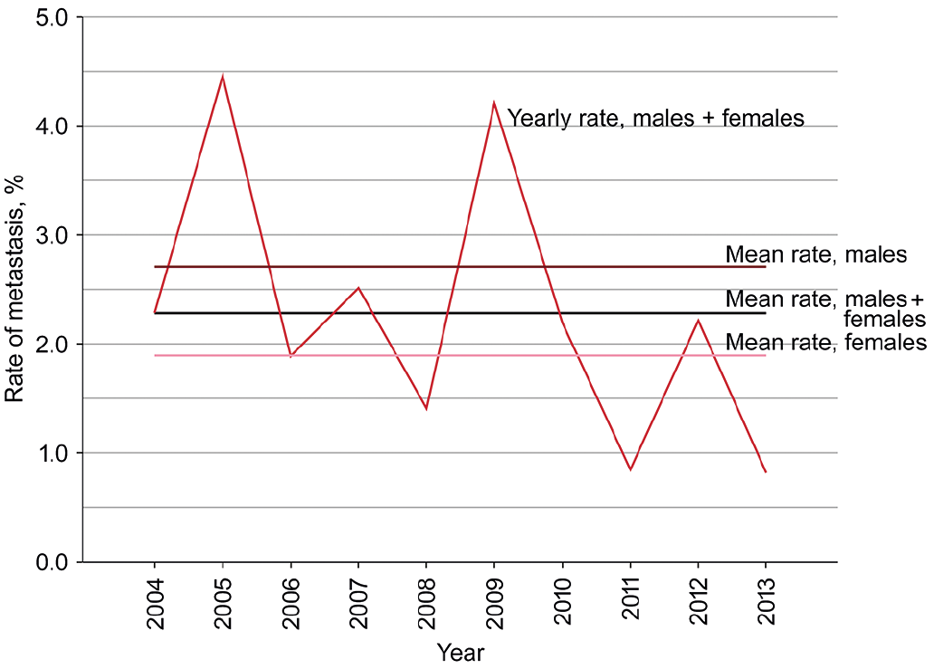 Click to show fullsize
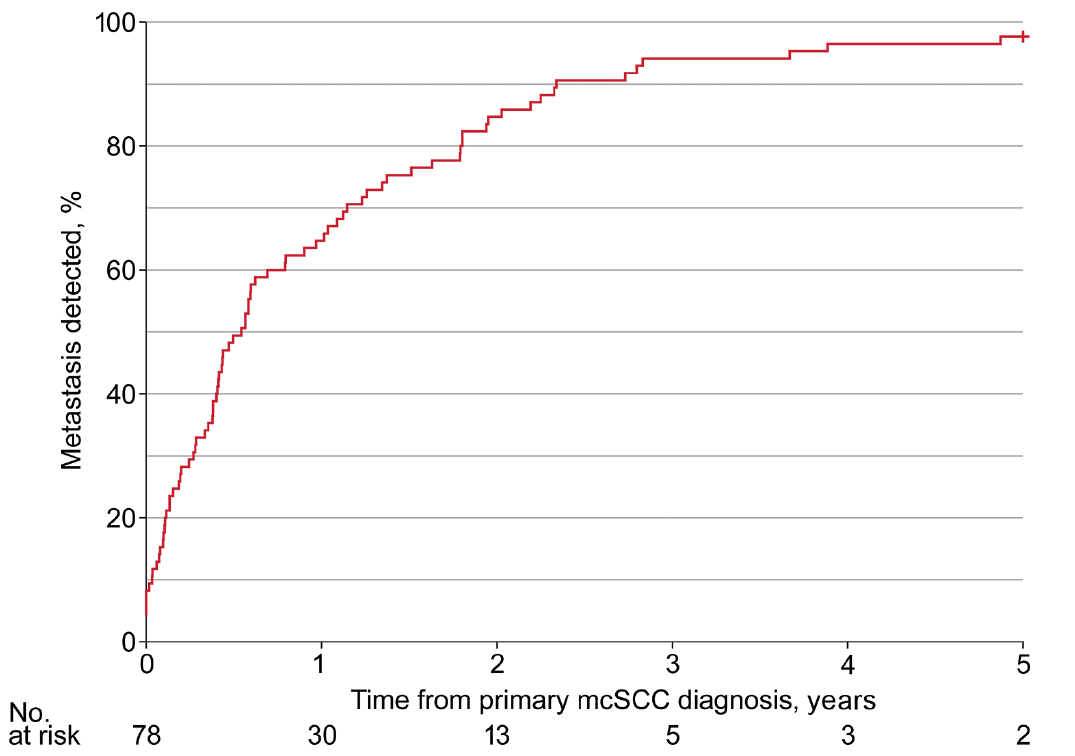 Click to show fullsize
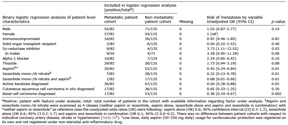 Click to show fullsize
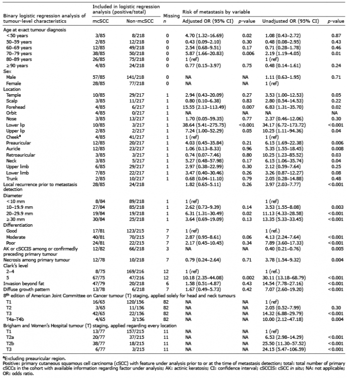 Click to show fullsize
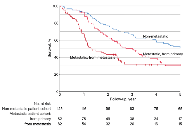 Click to show fullsize
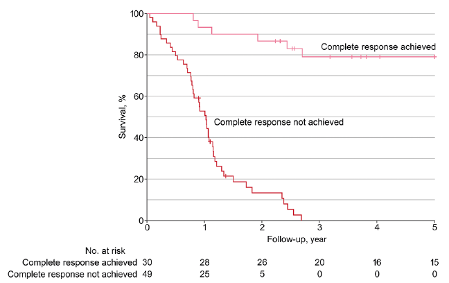 Click to show fullsize
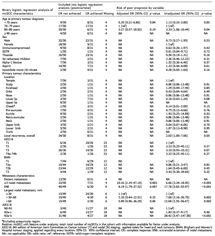 Click to show fullsize