


1University of Genoa, DiSSal Section of Dermatology, and 2Section of Pathology, University of Genoa, San Martino Polyclinic Hospital IRCCS, Largo R. Benzi 10, IT-16132 Genoa, Italy. E-mail: emanuele.cozzani@unige.it
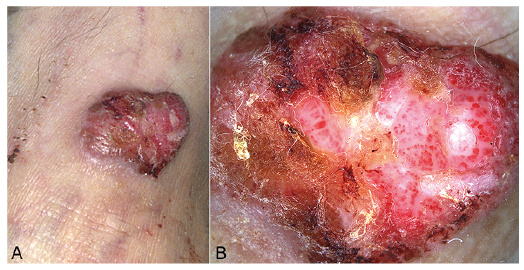
A 70-year-old Caucasian woman presented with a 40-year history of an asymptomatic erythematous, ulcerated nodule on her left ankle (Fig. 1A). The lesion had first appeared as a red, dome-shaped papule when she was 30 years old. Since then it had slowly enlarged and recently ulcerated.
Physical examination revealed a 40×30 mm, soft, ery-thematous, partially crusted, moist-appearing nodule, with a wafer-like scale collarette. Minor trauma made the lesion bleed (Fig. 1A). Her medical history included a previous non-Hodgkin’s lymphoma. The woman was generally in good health.
Dermoscopy revealed glomerular blood vessels and red clods in a reticular and curvilinear pattern, resembling a pearl necklace, with combined thin and thick white intersecting lines (Fig. 1B).
A skin biopsy showed well-defined acanthosis with regular psoriasiform hyperplasia, rete ridges of varying width, and partial fusion of rete ridges. The tumour cells comprised pale staining keratinocytes, replete with intracellular glycogen, staining positive with periodic acid-Schiff (PAS) (Fig. 2).
What is your diagnosis? See next page for answer.
Fig. 1. Clinical presentation. (A) Erythematous, ulcerated, partially crusted and partially moist appearing nodule measuring 40×30 mm, with a wafer-like scale collarette. (B) Dermoscopic appearance with linear serpiginous large glomerular vessels and red clods organized in a “string of pearls” pattern and thin and thick white intersecting curved lines.
Fig. 2. (a) An epidermal acantholytic area with proliferation of clear cells. Note the abrupt transition to and from the normal epidermis. The lesion shows abundant intracytoplasmic glycogen, Periodic acid-Schiff (PAS) positive, possibly removed by digestion with diastase. PAS stain (2×). (b) The epidermis is characterized by psoriasiform acanthosis, fusion of rete ridges, hypogranulosis and keratinocytes containing pale eosinophil cytoplasm with bland cytological features. Neutrophilic exocytosis, parakeratotic micro-abscesses and dilated blood vessels are present in the upper dermis. Haematoxylin and eosin (H&E stain) (20×).
Acta Derm Venereol 2020; 100: adv00288.
Diagnosis: Giant clear cell acanthoma
Clear cell acanthoma (CCA), also known as “Degos acan-thoma” was first described by Degos et al. in 1962 (1). It is a rare benign tumour with unknown aetiology. The tumour is probably of epidermal origin, though pilar and sweat gland origins are also debated (2). Some even argue that CCA represents a reactive inflammatory dermatosis, as immunohistochemical staining shows increased expression of cytokeratins, similarly to other inflammatory dermatoses, such as psoriasis (2). CCA is most commonly located on the lower extremities, as in our patient, but it can also present on the upper extremities, trunk, face, inguinal area, and CCA cases of the nipple and areola have also been described (3).
Clinically, CCA typically presents as a dome-shaped erythematous, asymptomatic papule or nodule, with a stuck-on appearance and a characteristic collarette of scale, classically described as “wafer-like” (2). The surface is either moist or crusted (4) and the lesion enlarges slowly over several years and ranges in size from 3 to 20 mm (4). Several clinical variants of CCA have been described, includ-ing a giant, pedunculated or polypoid, eruptive, pigmented, atypical and cystic variant (4).
The giant type measures > 4 cm, and only 9 cases of giant CCA have been reported in literature (5–12). The clinical characteristics of all 9 cases are shown in Table I. The current case would be the 10th case of giant CCA.
Dermoscopy is extremely characteristic, showing a linear serpiginous “string of pearls” vascular pattern that is highly suggestive for diagnosing CCA (3).
Regarding vascular findings in mature CCA, vessels acquire a more coiled appearance, while maintaining their typical serpiginous arrangement. In fact, as already suggested by Iseki et al. (13), mature CCA lesions, such as the one in the current case, as well as early CCA lesions, exhibit the typical vascular string of pearls formation. This makes diagnosis by dermoscopy relatively easy, even in longstanding, eventually ulcerated lesions. In fact, we confirm that regardless of CCA maturity, the dermoscopic “string of pearls” vascular pattern, reflecting large glomerular vessels and red clods, is a highly suggestive diagnostic clue to mature CCA. Furthermore, thin and thick white intersecting curved lines, as proposed by Iseki et al. (13) as a typical sign of mature CCA, were also observed in the current patient (Fig. 1B).
The primary clinical and pathological differential diag-noses that must be considered include eccrine poroma, basal cell carcinoma, squamous cell carcinoma, amelanotic melanoma, pyogenic granuloma, lichenoid keratosis, inflamed seborrheic keratosis and psoriasis. Among these lesions, however, the unique dermoscopic vascular pattern presented by CCA, as discussed previously, is highly suggestive for the right clinical-dermoscopic diagnosis.
Gold standard treatments for CCA are surgical excision or, provided that a biopsy confirmed the diagnosis, physical ablation with CO2 laser or liquid nitrogen. A conservative approach with topical calcipotriol 0.005% cream has also been reported to lead to complete resolution, which could be considered, especially for giant lesions or elderly patients (14).
Table I. Giant clear cell acanthoma cases reported in literature


 Click to show fullsize
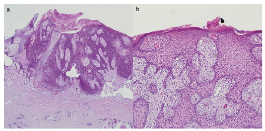 Click to show fullsize
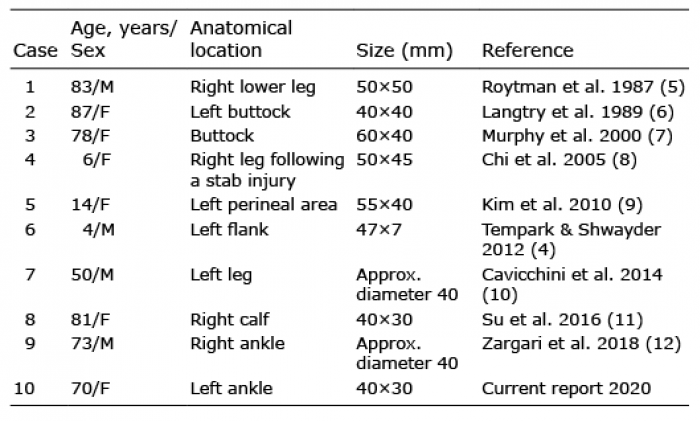 Click to show fullsize