


1Department of Dermatology, Faculty of Medicine, Academic Assembly, University of Toyama, 2630 Sugitani, Toyama 930-0194 and 2Department of Pharmacology, Faculty of Medicine, Kansai Medical University, Osaka, Japan. E-mail: tmakino@med.u-toyama.ac.jp and nakamtom@hirakata.kmu.ac.jp
Accepted Dec 22, 2020; Epub ahead of print Dec 29, 2020
Acta Derm Venereol 2021; 101: adv00372.
doi: 10.2340/00015555-3738
Solar elastosis is the accumulation of disorganized and non-functional elastotic material, which is found in photo-aged skin (1). Solar elastosis occurs through a cycle of elastic fibre degradation, followed by extracellular matrix production and reassembly into an organization that differs from the original structure. The exact pathomechanism of solar elastosis remains unknown; however, it has been reported to be associated with matrix metalloprotease 12 in elastin degradation (2) or ultraviolet (UV) radiation-induced alternative splicing of the ELN gene (3). Elastic fibres consist of microfibrils and polymerized elastin. Microfibrils are composed of homopolymers of fibrillin-1 and -2 together with latent transforming growth factor beta (TGF-β) binding proteins (LTBPs) and microfibril- associated glycoproteins (4). LTBPs are extracellular matrix proteins that share structural homology with fibrillins (5). Among the 4 LTBPs, LTBP-4 promotes elastin deposition onto microfibrils through interaction with fibulin-5, which is a tropoelastin-binding protein (6). LTBP-2 also interacts with fibulin-5, and directs fibulin-5 to deposit on fibrillin-1 microfibrils (7). Moreover, fibulin-4 conducts elastogenesis via interaction with the cross-linking enzyme lysyl oxidase (8). This study examined the expression of these elastogenic factors in solar elastosis to clarify its pathomechanism.
Human skin tissue samples with solar elastosis were obtained from elderly individuals (patient numbers 1–8 in Table SI; mean age 76.0 years) at Toyama University Hospital, Toyama, and Kansai Medical University, Osaka, Japan. The diagnosis of solar elastosis was made based on histological findings by dermatologists and pathologists. Sun-exposed aged skin without solar elastosis (patient numbers 9–15; mean age 72.1 years), sun-protected aged skin (patient numbers 16–26; mean age 73.5 years) and young skin (patient numbers 27–31; mean age 22.0 years) were obtained for use as controls. Detailed information on the individuals is shown in Table SI. All subjects provided written informed consent, and the study protocol complied with the principles of the Declaration of Helsinki. This study was approved by the medical ethics committees of the University of Toyama and Kansai Medical University. Immunofluorescence was performed as described previously (6). Mouse anti-elastin monoclonal antibodies (Merck, Dermstadt, Germany) and rabbit anti-human fibrillin-1 polyclonal antibodies (Elastin Products Co., Inc., Owensville, USA) were used as primary antibodies. Mouse anti-fibulin-4 monoclonal, mouse anti-fibulin-5 monoclonal, chicken anti-LTBP-2 polyclonal, and rabbit anti-LTBP-4 polyclonal antibodies were produced as described previously (6–8).
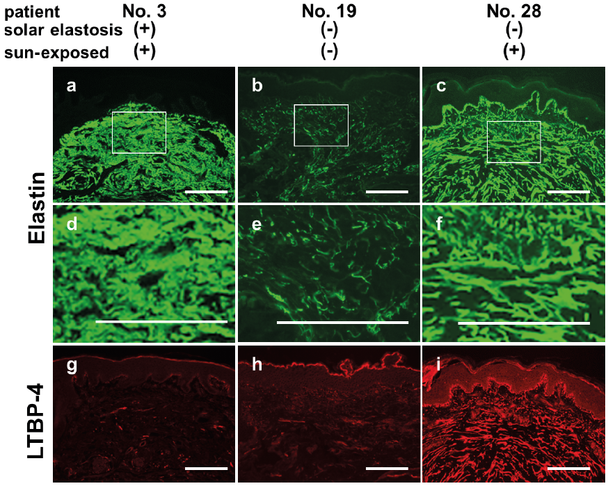
In sun-protected aged skins and sun-exposed, non-solar elastotic aged skins, dermal elastic fibres were degraded, and elastin deposition on microfibrils was decreased in comparison with young skin (Fig. 1b, e; Fig. S1). The oxytalan fibres (i.e. microfibril bundles extended from the lamina densa into the papillary dermis) were also degraded in 1 of 7 sun-exposed, non-solar elastotic aged skin samples and in 5 of 11 sun-protected aged skin samples. In contrast, in solar elastotic skin, intense staining of elastin was observed in a thick structure in the dermis, although oxytalan fibres were greatly reduced in 7 out of 8 samples (Fig. 1a, d). In most of the tissue samples with solar elastotic skin, intense staining of fibrillin-1, LTBP-2, and fibulin-4 was co-localized with the thick dermal structure, which was positive for elastin, although the fibrillin-1 signals showed a fragmented pattern in 2 of 8 cases (Fig. S1). The expression of these proteins decreased in both the dermal elastic fibres and the oxytalan fibres of aged skin samples without solar elastosis, in comparison with young skin. Notably, the expression of LTBP-4 was abolished or largely decreased, not only in non-solar elastotic aged skin samples, but also in solar elastotic skin, in comparison with young skin (Fig. 1g–i). All tissues of sun-exposed, non-solar elastotic aged skin samples and 4 out of 6 sun-protected aged skin samples showed decreased expression of fibulin-5 in dermal elastic fibres in comparison with young skin. In solar elastotic skin samples, 5 out of 8 samples showed intense fibulin-5 signals that were co-localized with elastin, while the rest of the samples showed decreased fibulin-5 staining (Fig. S1).
Fig. 1. Expression of elastic fibre components in solar elastotic skin. Immunofluorescent staining of (a–f) elastin and (g–i) LTBP-4 in (a, d, g) solar elastotic skin, (b, e, h) sun-protected aged skin, and (c, f, i) young skin. Boxes indicate the enlarged areas shown at the bottom of the figure. The results are representative of each group (solar elastotic skin: patient number 3; sun-protected aged skin: patient number 19; young skin: patient number 28). Scale bar: 100 µm for all panels.
In this study, elastin formed a thick layer in the upper dermis of solar elastotic skin, and intense staining of fibllirin-1, LTBP-2 and fibulin-4 was co-localized with elastin staining, although the expression of these proteins normally decreases with age in non-solar elastotic skin. It was reported that UV irradiation induces activation of elastin promoter (10). However, the formation of elastic fibre requires not only elastin, but also a series of elastogenic factors (11). Among these factors, we previously showed that LTBP-4 and fibulin-5 are necessary for the linear deposition of elastin onto microfibrils, and the loss of LTBP-4 causes formation of misplaced aggregation of elastin (6). Thus, it is of particular interest that the expression of LTBP-4 was largely diminished in all aged skin samples and was not re-induced in solar elastotic skin. These observations support a model for the pathogenesis of solar elastosis, in which ageing-related loss of LTBP-4 expression and UV-induced overexpression of elastin cause the accumulation of degenerated elastotic material in the upper dermis. Kadoya et al. reported decreased expression of fibulin-5 in aged skin and overexpression of fibulin-5 in a sample of solar elastotic skin (12). However, we found that 3 of 8 cases with solar elastosis showed decreased expression of fibulin-5, whereas the other cases showed increased expression of fibulin-5. These observations suggest that there are at least 2 types of solar elastosis; one with increased fibulin-5 and the other with decreased fibulin-5. Further study is needed to address whether fibulin-5 is involved in the development of solar elastosis. Nonetheless, the present study demonstrated that increased expression of fibrillin-1, LTBP-2 and fibulin-4 in combination with decreased LTBP-4 expression is associated with the development of solar elastosis. The largely diminished LTBP-4 expression in aged skin may also partly explain why elastogenesis is generally abrogated in aged skin.
This work was supported by Japan Society for the Promotion of Science (17K19534, 19H03439) and the Science Research Promotion Fund from the Promotion and Mutual Aid Corporation for Private Schools of Japan to TN.
The authors have no conflicts of interest to declare.


 Click to show fullsize