


1Center for Infectious Diseases, Queen Astrid Military Hospital, Rue Bruyn 1, BE-1120 Brussels, 2Travel Clinic, Institute of Tropical Medicine, Antwerp, 3General Internal Medicine, University Hospitals Leuven, Leuven, 4Inter-regional University Center of Expertise in Hospital Histopathology, Jumet, 5Department of Histopathology, Erasme University Hospital, Brussels, Belgium, 6Department of Cardiology, Erasmus Medical Center, Rotterdam, The Netherlands and 7Department of Clinical Sciences, Institute of Tropical Medicine, Antwerp, Belgium. E-mail: Isabella.strachinaru@mil.be
Accepted Feb 23, 2021; Epub ahead of print Mar 9, 2021
Acta Derm Venereol 2021; 101: adv00414.
doi: 10.2340/00015555-3777
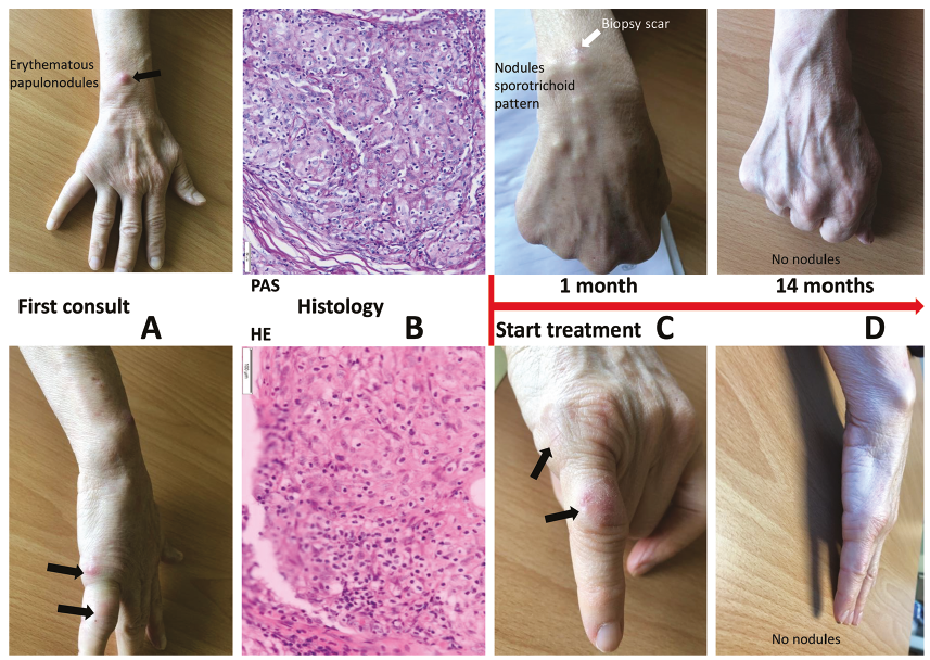
An immunocompetent 60-year-old woman presented to an outpatient clinic with nodular lesions on her right arm after getting stung in the 5th finger by a plant thorn while gardening. Inflammation appeared at the laceration site, and in the following weeks several subcutaneous nodules spread along a lymphatic trajectory, despite initial empirical treatment with amoxicillin followed by flucloxacillin. Her medical history was unremarkable. She owned a tropical fish tank, which she cleaned without any protection, and also owned 2 cats. There was no recent history of travel. She reported no fever or systemic symptoms. On examination, erythematous papulonodules (Fig. 1A) spanned from her 5th finger to her elbow in a sporotrichoid pattern. The rest of the clinical examination was unremarkable. All laboratory tests were within normal ranges or negative. Soft-tissue ultrasound revealed a 2-mm foreign object at the injury site and several hypoechoic and hypervascular nodules in the dermis, but no deep tissue involvement, abscesses or tenosynovitis. Histopathology of a nodule demonstrated dermal granulomatous inflammation with multinucleate giant cells, lymphocytes, histiocytes and neutrophils. Periodic acid-Schiff (PAS) and Fite stains were negative. No acid-fast bacilli were detected on direct Ziehl-Neelsen (ZN) staining. Bacteriological cultures, incubated at 30°C and 37°C, were negative. Based on the hobby-related exposure, M. marinum infection was evoked. However, diagnostic tests remained inconclusive and new lesions appeared, therefore the foreign object (a fragment of a thorn) was removed and another nodule was biopsied. The biopsy revealed massive granulomatous infiltration of the dermis with multinucleate giant cells and histiocytes, but no neutrophils (Fig. 1B). Some granulomas had central fibrinoid necrosis. PAS, Grocott and ZN stains and a immunohistochemical search for Mycobacterium tuberculosis were negative. Tissue cultures, incubated on blood agar plates, Sabouraud medium and mycobacterium-specific media at 30°C and 37°C, were negative after 3 months. As the diagnosis remained elusive, the patient underwent a third biopsy. In addition to previous tests, PCR for 16S rRNA, M. tuberculosis, Aspergillus and panfungal were performed. Although no antibiotics had been prescribed during the diagnostic process, all tests were negative. The patient reported that her fish had developed nodules on their skin and several had died; therefore, the aquarium water was cultured. One sample grew M. marinum, and another sample Mycobacterium alsense, another slow-growing NTM. M. alsense has been isolated from human respiratory samples (8), but no skin or soft-tissue involvement has been reported. Given the typical presentation in both patient and fish, M. marinum was considered responsible. Oral clarithromycin, 1 g/day, was started 173 days after the first consultation, and continued for 14 months due to slow remission of the lesions, typical of M. marinum infections with sporotrichoid pattern (Fig. 1C, D). The therapy was discontinued 1 month after the lesions subsided. A follow-up 1 month later showed no lesions. Another visit is scheduled 6 months after treatment discontinuation, according to experts’ recommendations (9).
Fig. 1. (A) first clinical examination. (B) Periodic acid-Schiff (PAS) stain, 20× magnification, shows no fungal element; haematoxylin-eosin stain, 10× magnification, shows epithelioid granulomas in the dermis. (C) Follow-up at 1 month of treatment. (D) Follow-up at 14 months of treatment.
This case highlights the diagnostic challenges that M. marinum poses, even when suspicion is high. In this case, adequate treatment was initiated more than 6 months after the first nodule appeared. In Holden’s series, the median time from presentation of symptoms to diagnosis was 194 days (maximum 548 days) (10), while Johnson reported a median time of 3.5 months, with extremes of up to 24 months (6). This case also illustrates how a broad diagnostic approach enabled M. marinum to be singled out from other infectious (11) and non-infectious diseases, such as neoplasms or reactive dermatoses, which can also present with nodular lesions distributed in a sporotrichoid pattern. A history of exposure to fish can raise suspicion of M. marinum infection, but histology and cultures or positive PCR are needed to confirm the diagnosis. In M. marinum infection, histopathology reveals granulomatous inflammation without caseation. Fibrinoid necrosis may be present in the granulomas, whilst lymphohistiocytic infiltrates and Langhans giant cells are present in the dermis (5, 12, 13). Early lesions usually present as conglomerates of polymorphonuclear cells surrounded by histiocytes (12). M. marinum may be identified on ZN or Fite stains, but its absence does not exclude diagnosis, as few bacilli are present in the lesions, except in immunocompromised patients (5, 13, 14). The current gold standard for diagnosis is mycobacterial culture. However, cultures are positive in only 40–60% of cases (5, 15). Clinicians suspecting M. marinum infection should therefore notify the laboratory of the suspicion. Specimens should be delivered on ice if the transport time to the laboratory is longer than 2 h and then be cultured on selective media. M. marinum grows optimally at 30°C, but poorly at 37°C (1). Cultures should be kept for 6 weeks before being reported negative (2). Failure to observe these conditions may result in false-negative cultures. PCR can improve the diagnostic yield of cultures, but is not 100% sensitive. In Sia’s series, Mycobacterium 16S rRNA PCR was positive in only 46% of cases, but 60% of the culture-negative specimens were positive by PCR, which highlights the usefulness of molecular techniques (5).
In selected challenging cases when M. marinum is suspected, the diagnostic approach should include aquarium water and fish cultures, alongside histological investigations, mycobacterial culture, and 16S rRNA PCR. At times, this may be the only method that can be used to reach a diagnosis.
The patient provided written consent for publication of this case report and the accompanying images. A copy of the written consent is available for review.
The authors have no conflicts of interest to declare.


 Click to show fullsize