


1Department of Dermatology, Tampere University Hospital, 2Celiac Disease Research Center, Faculty of Medicine and Health Technology, Tampere University, 3Department of Internal Medicine, Tampere University Hospital, 4Faculty of Social Sciences, Tampere University and 5Department of Pathology, Fimlab Laboratories, Tampere, Finland
Dermatitis herpetiformis is a cutaneous manifestation of coeliac disease. Anaemia is a common finding in patients with untreated coeliac disease, but little is known about the occurrence of anaemia in those with dermatitis herpetiformis. This study investigated the prevalence of anaemia and factors associated with anaemia in 250 patients with dermatitis herpetiformis, at diagnosis and one year after diagnosis. As controls, 139 patients with coeliac disease were included. Patient records were reviewed to gather baseline clinical, histological, and laboratory data. Follow-up data for patients with dermatitis herpetiformis were collect-ed from patient records and via questionnaires or at follow-up visits. The prevalence of anaemia was 12% in patients with dermatitis herpetiformis and 17% in patients with coeliac disease at diagnosis (p = 0.257). Anaemia in patients with dermatitis herpetiformis was not associated with the severity of skin symptoms or small bowel damage. The prevalence of anaemia at a 1-year follow-up had increased to 19%, but it was associated mainly with dapsone treatment.
Key words: coeliac disease; dapsone; gluten-free diet; villous atrophy.
Accepted Apr 1, 2021; Epub ahead of print Apr 13, 2021
Acta Derm Venereol 2021; 101: adv00443.
doi: 10.2340/00015555-3795
Corr: Teea Salmi, Department of Dermatology, Tampere University Hospital, FIN-33521 Tampere, Finland. E-mail: teea.salmi@tuni.fi
Dermatitis herpetiformis is a cutaneous manifestation of coeliac disease. Anaemia is a relatively common finding in patients with untreated coeliac disease, but little is known about the prevalence of anaemia in patients with dermatitis herpetiformis. This study showed that the prevalence of anaemia at diagnosis did not differ significantly between 250 patients with dermatitis herpetiformis and 139 patients with coeliac disease. Anaemia at diagnosis in dermatitis herpetiformis was not associated with the severity of skin symptoms or small bowel damage. The prevalence of anaemia at a 1-year follow-up had increased slightly, but it was associated mainly with dapsone treatment, which is generally known to have potential haematological side-effects.
Dermatitis herpetiformis (DH) is a blistering and itching skin disease and a cutaneous manifestation of coeliac disease (1). In DH and coeliac disease, digested gluten provokes a wide spectrum of clinical symptoms in genetically susceptible individuals (2). Although patients with DH rarely develop prominent gastrointestinal symptoms, three-quarters of them have a variable degree of small bowel mucosal villous atrophy (3, 4), and those with normal villous architecture evince intestinal coeliac-type inflammation (5, 6). Furthermore, characteristic of both DH and coeliac disease, is an autoantibody response targeted against tissue transglutaminase (TG2) (7, 8). A pathognomonic feature of DH, however, is the presence of granular IgA deposits in the dermis, directed against epidermal transglutaminase (TG3) (9).
Strict lifelong adherence to a gluten-free diet (GFD) is an effective treatment for DH and coeliac disease. The diet alleviates the gastrointestinal and, also eventually, cutaneous symptoms, and results in normalization of the small bowel mucosal architecture (10, 11). In addition, dapsone medication is initiated for most patients with DH to quickly alleviate the troublesome skin symptoms (12).
The clinical picture of coeliac disease has changed over time, and the frequency of classical coeliac disease symptoms, such as chronic diarrhoea and weight loss, has decreased, while, concomitantly, non-classical manifestations have become more common (13). Anaemia is one of the most common presentations of coeliac disease (13, 14), occurring even as the only manifestation of an otherwise silent coeliac disease (15). In studies performed in Western countries, the prevalence of anaemia among people with untreated coeliac disease is in the region of 20–30% (16–19). Iron deficiency anaemia seems to be the most common form of anaemia in coeliac disease, but vitamin B12 and folate deficiencies, as well as anaemia of chronic disease, also occur (17, 20). The main, but not the sole, mechanism for anaemia in coeliac disease is thought to be malabsorption caused by the damaged small bowel mucosa (14, 17, 20), and in the majority of studies, anaemia has been associated with more severe villous atrophy and higher autoantibody levels at diagnosis (18, 19, 21, 22).
There is scant knowledge regarding anaemia in patients with DH. A few earlier studies have shown that the prevalence of anaemia may be as high as 42% among patients with treated DH (23, 24). However, even though it is frequently used in the initial treatment in addition to a GFD, dapsone has a well-known adverse effect of dose-dependent haemolysis (25). To our knowledge, only one study has investigated the frequency of anaemia among patients with untreated DH, finding the prevalence of anaemia to be 23% (26).
The aim of the current study was to examine the prevalence of anaemia in a large well-defined cohort of patients with untreated DH and to compare it to anaemia in patients with coeliac disease. A further aim was to identify the factors associated with anaemia at diagnosis of DH and after adherence for one year to dapsone and/or GFD treatment.
Patients and controls
This study included 250 adult patients with DH (≥ 18 years of age) with an available blood haemoglobin (Hb) value investigated at the time of diagnosis of DH. The patients had been diagnosed in 1970–2019 at a special outpatient clinic for patients with DH at the Department of Dermatology, Tampere University Hospital, Tampere, Finland, and for each patient, the diagnosis of DH was based on the typical clinical picture and the demonstration of dermal granular IgA deposits in perilesional skin immunofluorescence biopsies. After diagnosis, all patients were routinely referred to undergo gastroscopy with small bowel biopsies, and were further advised to adhere to a strict, lifelong GFD.
Dapsone medication was initiated, according to routine practice, for patients with DH who had severe skin symptoms. All patients were followed-up at a special DH outpatient clinic until the DH rash had resolved and the dapsone medication could be discontinued.
As a control group, 139 patients with coeliac disease diagnosed at ≥ 18 years of age and with an available diagnostic Hb value were included. All patients had a small bowel biopsy-confirmed diagnosis of coeliac disease performed at Tampere University Hospital during the same time period as the patients with DH, and all were considered to have the classical presentation of coeliac disease.
Study protocol
The medical records of all participating patients with DH and with coeliac disease, from the time of the diagnosis, were reviewed. Data on demographic characteristics, presence of gastrointestinal symptoms, small bowel mucosal findings, serum coeliac autoantibody tests, and Hb levels were gathered. The small bowel mucosal histology was interpreted by an experienced pathologist, and the result was graded as subtotal or total villous atrophy, partial villous atrophy, or normal mucosa, as described previously (3). The serum IgA class coeliac disease autoantibody tests were anti-reticulin antibody (ARA), endomysium antibody (EmA), or TG2 antibody tests, depending on the time of testing.
In addition, data on the duration of skin symptoms before diagnosis, severity of skin symptoms at diagnosis, initiation of dapsone treatment, and levels of serum vitamin B12 or transcobalamin II-bound vitamin B12 (B12-TC2), as well as erythrocyte folate (E-folate) or serum folate (S-folate) were collected from the medical records of all study patients with DH. Serum ferritin and transferrin receptor test results were also recorded, but since they are only sparsely investigated in DH, their availability from medical records was very low, and these parameters were excluded from further analysis. The skin symptoms were graded as mild, moderate, or severe by a dermatologist, and the grading was based on the presence of a few, several, or many blisters, macular eruptions, and erosions on the knees, elbows, buttocks, scalp, or elsewhere on the body
For patients with DH, the 1-year follow-up data on the positivity of serum coeliac autoantibody tests and Hb levels were recorded from the patient records. Follow-up data on the duration of skin and gastrointestinal symptoms, as well as the duration of dapsone treatment, were gathered from DH-specific questionnaires sent to all patients with DH diagnosed between 1970 and 2014, as describ-ed previously (27), and from the remainder at follow-up visits to the DH outpatient clinic. The DH-specific questionnaire included both open and multiple-choice questions, and even though it is not validated, it has been used previously in several studies on DH (27, 28). The duration of skin symptoms after diagnosis was further classified into 3 groups: less than one year, 1–2 years, and longer than 2 years. For those receiving dapsone after diagnosis, the duration of dapsone treatment was interpreted as the duration of skin symptoms, since dapsone is routinely discontinued as soon as the skin symptoms are controlled with a GFD alone. For those not using dapsone, the duration of skin symptoms was recorded from the questionnaire. Moreover, the duration of gastrointestinal symptoms after diagnosis was classified as: no symptoms, less than 3 months, 3–12 months, and over 12 months.
The Regional Ethics Committee of Tampere University approv-ed the study protocol and usage of the register-based data. All subjects provided written informed consent.
Laboratory parameters
The following laboratory values were measured by standard laboratory methods: blood Hb (reference values: 117–155 g/l for women and 134–167g/l for men), serum vitamin B12 or B12-TC2 (reference values 145–570 pmol/l and > 35 pmol/l, respectively) and E- or S-folate (reference values 1,187–2,854 nmol/l and 8.8–42.4 nmol/l, respectively).
In the ARA and EmA tests, titres 1:≥ 5 were considered positive. In the TG2 antibody tests, the reference value was 20 or 5 U/ml, depending on whether an INOVA (INOVA Diagnostics, San Diego, CA, USA) or Celikey (Celikey Pharmacia, Uppsala, Sweden) test was used. The ARA, EmA, and TG2 antibody tests are all directed against TG2 (29), and in this study, a positive result in any of these tests was interpreted as positive coeliac disease serology.
Statistical analysis
The patients with DH were divided into 2 groups based on whether they had anaemia or normal Hb, according to the reference values at the time of the diagnosis or at the 1-year follow-up. A change in Hb value ≥ 10 g/l during the first year of treatment was considered clinically relevant, and patients with DH were divided into 3 groups depending whether the Hb value had decreased, remained constant, or increased during the first year of treatment according to the clinically relevant criteria. Categorical variables were presented as numbers and percentages, and continuous variables as medians with quartiles (Q1–Q3) or ranges (min–max), as the majority of the data were skewed. A χ2 test or Fisher’s exact test was used in cross-tabulations, and the Mann–Whitney U test or Kruskal–Wallis H test was used for comparing continuous variables. All statistical analyses were performed using SPSS version 26. Statistical significance was set at p < 0.05 and testing was two-sided.
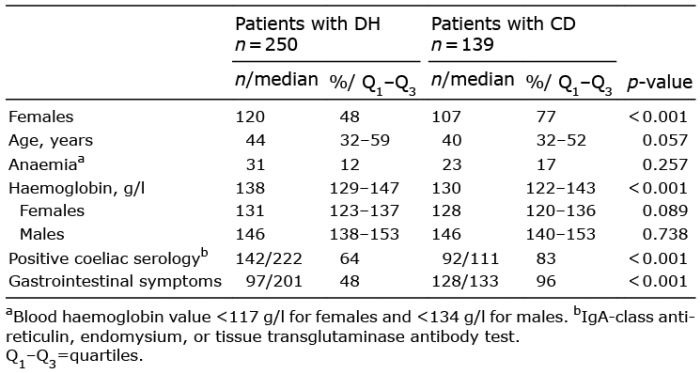
Table I. Demographic, clinical, and histological data and laboratory values of 250 patients with dermatitis herpetiformis (DH) and 139 control patients with coeliac disease (CD) at the time of diagnosis
When patients with DH with anaemia and those without anaemia at the time of the diagnosis were compared, patients with normal Hb more frequently had gastrointestinal symptoms at diagnosis (52% vs 18%, p = 0.003), and they also reported a statistically significantly longer duration of gastrointestinal symptoms after diagnosis compared with patients with DH with anaemia (p = 0.004) (Table II). Age, sex, severity and duration of skin symptoms before or after diagnosis, small bowel histology, and coeliac seropositivity at diagnosis did not differ between patients with DH with and without anaemia. In addition, the prevalence of vitamin B12 and folate deficiencies were similar between patients with and without anaemia.
Table II. Demographic, clinical, histological, and serological data of 31 patients with dermatitis herpetiformis (DH) with anaemiaa and 219 patients with DH with normal blood haemoglobin (Hb) values at the time of diagnosis
Among the patients with DH, anaemia was slightly more common in males compared with females, but the difference was not statistically significant (15% vs 9%, p = 0.136). Male patients with DH more frequently had severe skin symptoms at diagnosis compared with females (38% vs 25%, p = 0.022), and males also needed dapsone treatment more often than females did (84% vs 68%, p = 0.004). The presence of villous atrophy did not differ between male and female patients with DH (74% vs 73%, p = 0.924).
At time of follow-up
Of all patients with DH, 78% had adopted a GFD and 58% were on dapsone treatment at the 1-year follow-up. The prevalence of anaemia was 19% among 160 patients with DH, and in the patients with DH adhering to GFD and with available data (n = 76), the prevalence was higher among those using dapsone compared with those not using dapsone (28% vs 0%, p = 0.001). Also, anaemia prevalence was higher in males with DH compared with females with DH (25% vs 14%), although the difference did not reach statistical significance (p = 0.085). The median Hb value in males with DH had decreased significantly, from 146 to 141 g/l (Q1–Q3 = 135–149, p < 0.001) and in females non-significantly, from 131 to 128 g/l (Q1–Q3 = 121–137, p = 0.490).
Patients with DH who had anaemia at the 1-year follow-up more frequently had ongoing dapsone treatment compared with those with normal levels of Hb (80% vs 53%, p = 0.026), and dapsone was more often used among males than females (66% vs 47%, p = 0.015). In addition, patients with DH who had anaemia at follow-up had a significantly (p = 0.006) longer duration of skin symptoms after diagnosis than those without anaemia (>2 years, 65% vs 28%), but the duration of skin symptoms did not differ between the sexes. Otherwise, statistically significant differences were not detected between patients with DH with normal Hb and with anaemia at the follow-up in terms of age, adherence to GFD, duration or severity of skin symptoms at diagnosis, presence or duration of gastrointestinal symptoms before or after diagnosis, small bowel histology at diagnosis, and positivity of coeliac serology, either at diagnosis or at 1-year follow-up.
When patients with DH were further divided into 3 groups depending on whether the Hb had decreased, remained constant, or increased at the 1-year follow-up compared with the Hb level at diagnosis, 16% of the patients had decreased, 76% constant, and 8% increased Hb levels. Those with decreased Hb levels at follow-up had the highest median Hb levels at diagnosis (135 g/l in females and 155 g/l in males; p < 0.001) and those who had increased Hb at follow-up the lowest (median values at diagnosis 117 g/l in females and 125 g/l in males; p = 0.001 in both sexes). Patients with DH whose Hb had decreased during follow-up, had a longer duration of skin symptoms after diagnosis (p = 0.005) and also used dapsone more frequently at the 1-year follow-up compared with those with constant or increased Hb (p = 0.015) at follow-up, but no other significant differences were detected between the groups in demographic or clinical data, positivity of coeliac serology at diagnosis or follow-up, or bowel histology at diagnosis.
This study established that anaemia is a rather infrequent finding in untreated DH; only 12% of patients had anaemia at diagnosis. The presence of anaemia was not associated with the severity of skin symptoms or degree of small bowel villous atrophy. Although the prevalence of anaemia was slightly higher, at 17%, in untreated coeliac disease, in which small bowel damage is known to be more severe than in DH, the difference did not reach statistical significance in the current study. A relevant finding was that clinical recovery was equal in patients with DH with and without anaemia when diagnosed, as the duration of skin symptoms was similar and gastro-intestinal symptoms alleviated even more rapidly among those with anaemia at diagnosis. At the 1-year follow up, the prevalence of anaemia had increased to 19%, but this appeared to be associated mainly with prolonged skin symptoms and ongoing medication with dapsone.
The prevalence of anaemia at diagnosis in the current study was lower than that in the only previously perform-ed study, which investigated 65 patients with untreated DH in India and reported a 23% prevalence of anaemia (26). In patients with coeliac disease, in which anaemia has been far more extensively studied than in DH, the prevalence of anaemia in untreated coeliac disease has been reported to be slightly higher (23–40%) in European studies (16, 19, 22, 30) compared with the current study. However, the prevalence of anaemia in patients with coeliac disease has been reported to be as high as 93% in India (20), and thus the prevalence of anaemia appears to be highly dependent on geographical location. Different geographical locations could therefore offer a possible explanation for the lower prevalence of anaemia in the current study compared with the previous Indian study of DH (26).
The current study also analysed the factors associated with anaemia at diagnosis of DH, and found that anaemia was not associated with the severity of skin symptoms and, rather surprisingly, that the patients without anaemia had gastrointestinal symptoms more frequently than those with anaemia. The results of previous studies of coeliac disease are conflicting regarding the association of anaemia and gastrointestinal symptoms (19, 31, 32), and current evidence thus suggests that clinical symptoms are not reliable markers of anaemia in coeliac disease or DH. In addition, in the current study, small bowel mucosal damage, or the presence of serum coeliac autoantibodies was also not associated with anaemia. In coeliac disease, some studies have shown an association between anaemia and more severe villous atrophy and higher levels of serum TG2-antibody (18, 21, 22), whereas the opposite results also exist (33) in line with the current study of DH. This supports the suggestion that the aetiology of anaemia in coeliac disease and DH is multifactorial; even though malabsorption of nutrients due to small bowel damage exists, other aetiologies also contribute (17, 34). The multifactorial aetiology of anaemia is also suggested in the current study, as the majority of the patients with available results were shown to have vitamin B12 or folate deficiencies. This is a rather surprising finding, as vitamin B12 and folate deficiencies are known to be quite rare in coeliac disease; instead, iron deficiency is a much more common finding. The high prevalence of vitamin B12 or folate deficiencies in the current study may be due to selection bias, since vitamin B12 and folate values were not routinely tested in DH, but only in cases with a high pre-test likelihood of low values. Also, in the current study, we were unable to investigate iron parameters.
In the current study, the prevalence of anaemia at the 1-year follow-up, when most patients adhered to a GFD, was slightly higher than that in untreated DH. One obvious reason for this is dapsone medication, since 58% of the patients with DH used this drug at the 1-year follow-up, and a well-known side-effect of dapsone is dose-dependent haemolysis and anaemia (25). This is further supported by the facts that decreasing Hb values were significantly associated with the longer duration of skin symptoms after diagnosis and ongoing dapsone treat-ment, and anaemia prevalence was significantly high-er among GFD-treated patients with DH using dapsone compared with those not using dapsone. In addition, males had lower Hb values and males also used dapsone more frequently than did females at 1-year follow-up. There are scant previous studies of the occurrence of anaemia in patients with treated DH. In a study by Fry et al. (23) 5 out of 12 patients with DH undergoing dapsone treatment had anaemia. In another study from the UK (24), 86 patients with DH were investigated, of which 73% were on a GFD and 45% were on dapsone or sulphapyridine treatments. In that study, the total prevalence of anaemia was not reported, but 8 patients (8%) had macrocytic anaemia and, in 6 of these, the cause was probably dapsone treatment. One explanation for the rather low prevalence (19%) of anaemia among patients with treated DH in the current study may be the good adherence to a GFD in Finland. Even though the adherence to a GFD in the current study was relatively low (78%) at the 1-year follow-up, we have previously shown that, in Finland, in the long-term, 98% of our patients with DH adhere to a GFD (35). An additional explanation could be the recommendations for moderate dosage of dapsone in Finland. Dapsone is initiated in the majority of patients with DH (35) as a 25–50 mg daily dose, and it is increased up to 100 mg only if necessary.
Study strengths and limitations
The main strengths of the current study were the large, prospectively gathered, and carefully followed-up, series of patients with DH with a skin immunofluorescence biopsy-proven diagnosis and histologically confirmed coeliac disease control group. An additional strength was the availability of clinical, serological, and small bowel histological data for most patients with DH. Moreover, the study was performed in an area with a high prevalence of DH (36) and excellent GFD adherence rates (35). A limitation was that the study did not include healthy controls, and, in the follow-up, some information was based on questionnaires gathered retrospectively, which may have caused selection or recall bias. In addition, where was insufficient information on the weight, height, or iron status of the patients, and thus the current study was unable to thoroughly investigate the malabsorption status of the study patients. Furthermore, the dapsone doses were not counted.
Conclusion
This study found that the prevalence of anaemia in un-treated DH was rather low, at only 12%. The presence of anaemia was not associated with the severity of skin symptoms or villous atrophy at diagnosis, and it did not significantly differ between patients with DH and those with classical coeliac disease. Furthermore, the prevalence of anaemia in the dapsone and/or GFD-treated patients with DH increased to 19% at the 1-year follow-up. Decreased levels of Hb seemed to be confined mostly to those patients with DH who had ongoing skin symptoms and dapsone treatment.
This research was funded by the Academy of Finland, the Competitive State Research Financing of the Expert Responsibility area of Tampere University Hospital (grants 9X051 and 9AA070), Tampere University Hospital Support Foundation, the Finnish Coeliac Society, the Finnish Medical Foundation and the Finnish Dermatological Society.
The authors have no conflicts of interest to declare.


 Click to show fullsize
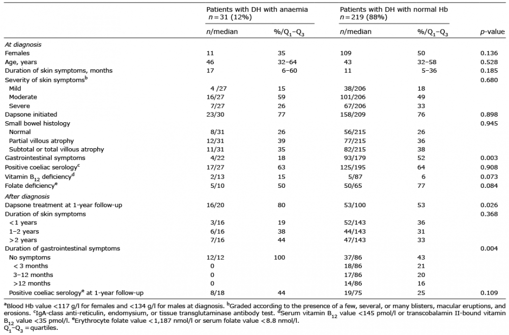 Click to show fullsize