


1Department of Dermatology and Copenhagen Wound Healing Center, 2Digestive Disease Center, Bispebjerg Hospital, University of Copenhagen, Nielsine Nielsens Vej 11, DK-2400 Copenhagen and 3Department of Clinical Medicine, Faculty of Health and Medical Sciences, University of Copenhagen, Copenhagen, Denmark. E-mail: Sven.Per.Magnus.Aagren@regionh.dk
Accepted May 10, 2021; Epub ahead of print May 18, 2021
Acta Derm Venereol 2021; 101: adv00465.
doi: 10.2340/00015555-3829
Zinc oxide (ZnO) is commonly applied to inflamed and injured skin due to the anti-phlogistic effects of bioactive Zn2+ (1, 2). Pretreatment of normal human epidermal keratinocytes (NHEKs) with Zn2+ reduced secretion of the proinflammatory cytokine interleukin (IL)-1α (3). Furthermore, topical administration of ZnO to murine skin increases keratinocyte mitosis (4), which is beneficial in wound healing. Non-invasive monitoring of inflammation and cell proliferation would provide extremely useful information. The aim of this randomized, double-blind, placebo-controlled trial (NCT03221699) was to study the anti-inflammatory and wound-healing effects of topical ZnO (1.1%) in an oil-in-water emulsion, by assessing skin erythema and complete wound closure in 2 standardized wound models. Furthermore, the usefulness of protein biomarkers, of inflammatory processes, IL-1α, and of epidermal proliferation, keratin 6A (K6A), were assessed (5, 6).
Participants, study design and flow, randomization, blinding and treatments have been described in our previous publication (7). In 30 Caucasian healthy volunteers, one axilla was randomized to daily application of ZnO and the contra-lateral axilla to daily application of placebo. After 8 days of treatment, duplicate lancet wounds (each 1.5-mm wide and 2.0-mm deep) were made 2 cm apart using a paediatric blood collection device (8) and 1 laser wound was made with an ablative fractional CO2 laser (7×7 mm and 0.8 mm deep) with a density of 10% and 20 mJ pulse energy (9) in the treated non-hairy axillary fossa. The daily treatments continued, and inflammation and wound healing were monitored by handyscope postwounding on days 3, 4 and 5. A scab/crust-free wound was considered completely healed. Surface skin proteins were collected with a 5.4-cm2 piece of adhesive tape (5) and analysed on IL-1α and K6A by enzyme-linked immunoassay (ELISA). The relevance of using K6A as biomarker of proliferation was investigated in proliferating vs stratifying NHEKs in vitro. Further details of the methods are given in Appendix S1.
The area under the erythema-time curve (AUC) was calculated and compared with the t-test (10). The IL-1α data were analysed by analysis of variance (ANOVA) and the K6A levels in NHEKs by the t-test. p < 0.05 was considered statistically significant.
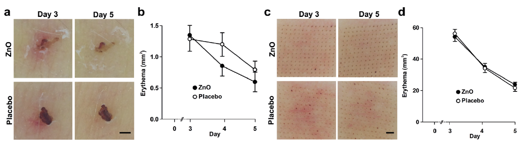
Erythema was slightly (p < 0.05) attenuated by topical application of ZnO around the incisional wounds, but not in laser-induced wounds (Fig. 1). The reason for the lack of effects of ZnO in laser-induced wounds on inflammation is unknown.
Fig. 1. (a, b) Erythema around lancet-induced wounds and (c, d) in laser wounds. (a, c) Images (20× magnification) were taken with a handyscope and (b, d) the erythematous areas (mm2) were quantified using ImageJ software (NIH, Bethesda, MD, USA). (b) The area under the curve (AUC) between days 3 and 5 decreased in zinc oxide (ZnO)-treated (1.81 ± 0.27 mm2 · day) lancet wounds (p < 0.05, 1-tailed paired t-test) compared with placebo-treated (2.23 ± 0.31 mm2 · day) wounds (14 wounds with ecchymosis were excluded). The mean of the areas of the anterior and posterior lancet wounds was used for statistical analyses. (d) The extent of erythema was similar for ZnO and placebo-treated laser wounds. Scale bar: 1 mm. Mean ± standard error of the mean (SEM).
On day 5, 16 of the 58 ZnO-treated lancet wounds were healed, compared with 18 of the 58 placebo-treated wounds. The corresponding healing rates for the laser wounds were 5 of 29 ZnO-treated and 4 of 29 placebo-treated wounds. That ZnO did not accelerate wound healing contrasts with previous findings (11). It is noteworthy that the ZnO formulation delivers at least 20-fold higher Zn2+ levels than is beneficial for epithelialization with ZnO incorporated in another carrier (7, 12). It is possible that the placebo alone promoted epithelialization due to its glycerol component, which increases skin hydration (13). MMP inhibitors delay epithelialization and could be included to obtain an idea of the range of detection (10).
Nyman et al. (14) demonstrated enhanced epithelialization of lancet wounds by intradermal injections of hyaluronic acid; 8 of 9 wounds on the forearm were completely epithelialized (as judged by histology) already on post-wounding day 1 compared with none in the control group. In addition, these authors found that erythema peaked on day 1 using polarized light spectroscopy (14), confirming previous findings with laser Doppler perfusion imaging (8). The current study found that digital photography with the handyscope, followed by image analysis, was convenient. The validity of images acquired with the handyscope for erythema measurements was confirmed using an open macro lens (Appendix S2).
The levels of the inflammation biomarker IL-1α were similar in the ZnO and placebo groups after 8 days of skin treatment and in the laser wounds on day 5, confirming the macroscopic erythema measurements (Fig. 2).
Fig. 2. Interleukin (IL)-1α in treated skin and day-5 laser wounds in the axilla. IL-1α was detected in 169 of the 176 tape-strip samples. IL-1α levels did not differ (p = 0.673) between the zinc oxide (ZnO) and placebo groups after 8 days of skin treatment or in the laser wounds (p = 0.599). Mean ± standard error of the mean (SEM).
This study searched for a biomarker of keratinocyte proliferation to reproduce the observed mitogenic effect of topical ZnO in the epidermis (4). The cytoskeletal protein K6 is associated with keratinocyte proliferation. However, K6A was undetectable in tape-strips of skin and treated laser wounds. Kinn et al. (6) sampled corneocytes from the forearms with chambers incubated with phosphate-buffered saline (PBS) for 30 min and reported K6 levels exceeding 1,000 pg/ml. This discrepancy was unlikely to be due to the ELISA assay, as the assay performed as expected in NHEKs, which showed increased (p = 0.003) intracellular K6A contents in proliferating (279 ± 46 pg/µg protein) vs differentiating (160 ± 26 pg/µg protein) NHEKs, or to extracellular K6A, which was not detected in concentrated (10×) conditioned medium. Arginase is elevated in psoriatic skin and has been suggested to reflect increased keratinocyte proliferation (15). In general, sampling of skin surface biomarkers by tape stripping is a very valuable method to elucidate physiological and pathophysiological mechanisms and mode of action of therapeutics and cosmetics on the skin.
The authors have no conflicts of interest to declare.


 Click to show fullsize
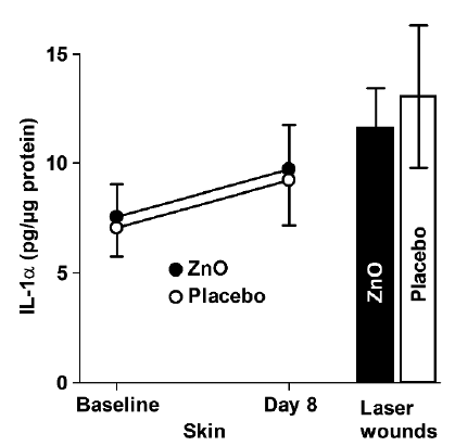 Click to show fullsize