


Department of Dermatology, Di.S.Sal., University of Genoa, San Martino Polyclinic Hospital IRCCS, Largo Rosanna Benzi 10, IT-16132 Genoa, Italy. E-mail: emanuele.cozzani@unige.it
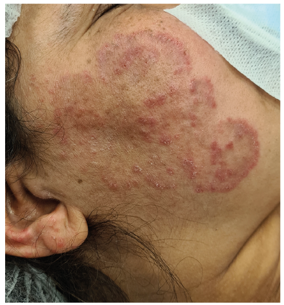
A 47-year-old Asian woman presented with pruritic, polycyclic-to-serpiginous, slowly migrating erythematous macules, with fine scaling and raised borders, on her cheek, ear and scalp, which had been present for 9 months (Fig. 1). She had had pulmonary, nodal mediastinal and uveal sarcoidosis for 2 years, which was initially treated with prednisone 50 mg/day, then tapered to 25 mg/day, adding methotrexate 10 mg/week. Her medical history was remarkable for gastric ulcer and reflux, as well as steroid-induced diabetes mellitus type 2, treated with gliclazide 30 mg/day.
Laboratory examinations, including antinuclear antibodies (ANA) and extractable nuclear antigens (ENA), were negative. The patient denied sun exposure in the past year.
A biopsy specimen was taken from the border of lesions, but histopathological examination with haematoxylin-eosin (HE) stain was non-specific.
What is your diagnosis? See next page for answer.
Fig. 1. Clinical image of the erythematous annular and pruritic macules, with slightly scaly and infiltrated borders on the cheek and ear of the patient.
Acta Derm Venereol 2021; 101: adv00531.
Diagnosis: Tinea incognita
Tinea incognita is a dermatophyte skin infection, promoted by erroneously administered topical or systemic corticosteroids, as well as immunosuppressants and diabetes mellitus. It lacks the typical manifestations of tinea, being less scaly, more pustular, irritated and extensive (1, 2). Immunosuppressors, such as steroids, reduce the cell-mediated inflammatory response, enabling the infection to spread, and modifying the clinical manifestation (1).
Because of its unusual appearance, tinea incognita is difficult to diagnose and should be suspected in cases of aspecific, erythematous, pruritic patches, in which the typical ringworm appearance of tinea is minimal (3).
In these cases, a reliable diagnosis is possible only by direct microscopical mycological examination on skin scrapings or histological slides, culture, or by PCR (1).
Indeed, Alcian blue (AB) stain and Periodic acid–Schiff (PAS) stain evidenced fungal hyphae in the stratum corneum and follicular plugs (Fig. 2), suggestive for mycotic skin infection. Notably, histological features of tinea can be subtle on HE stain; hence, PAS and AB staining may be needed to evidence fungal hyphae and make a diagnosis (4).
An additional scraping-test in the current case confirmed yeasts and moulds, which proved to be Candida guilliermondii, Fusarium solani and Fusarium oxysporum on PCR (5).
The patient received itraconazole 100 mg/twice daily for 2 weeks with complete remission. Oral antifungal therapy is necessary to cure tinea incognita. Itraconazole, at the above-mentioned posology, or 200 mg/twice daily for one week, is the treatment of choice (1).
As in the current case, tinea incognita may have unusual presentations, resembling gyrate, annular erythemas, such as annular cutaneous sarcoidosis (ACS), (6) erythema gyratum repens (EGR), (7) subacute cutaneous lupus erythematosus gyratus repens (SCLE-GR), (8) and annular elastolytic giant cell granuloma (AEGCG) (9).
In the current case, EGR was suspected due to the patient’s risk factors for gastro-oesophageal cancer (3, 7). EGR is, however, characterized by rapidly migrating lesions, and the patient had no anaemia, weight loss, or cancerous lesions on total-body tomography (7).
Specifically, ACS was suspected: skin involvement represents the second most frequent disease manifestation of sarcoidosis after pulmonary involvement (6). However, histological absence of non-caseating granulomas excluded cutaneous sarcoidosis (2). Also, AEGCG and SCLE-GR were excluded by histology, due to the absence, respectively, of granulomatous infiltrate with multinucleated giant cells (9) and SCLE features: epithelial atrophy, dermo–epidermal junction vacuolization and perivascular infiltrate (8).
The authors have no conflicts of interest to declare
Fig. 2. (A) Hyphae in the stratum corneum of the epidermidis (Alcian blue stain, original magnification ×200). (B) Keratotic follicular plug containing hyphae (Periodic acid-Schiff stain, original magnification ×40).


 Click to show fullsize
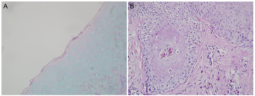 Click to show fullsize