


1Division of Dermatology, and 2Division of Cardiology, Niigata University Graduate School of Medical and Dental Sciences, Niigata, Japan. *E-mail: rh19840629@med.niigata-u.ac.jp
Accepted Jul 14, 2021; Epub ahead of print Jul 15, 2021
Acta Derm Venereol 2021; 101: adv00511.
doi: 10.2340/00015555-3881
Hydrophilic polymers are widely used coatings for cardiac catheterization and coronary angiography medical devices (1). These surface coatings have properties that enhance the biocompatibility and manoeuvrability of endovascular technologies, while decreasing friction and reducing trauma to the vessel walls (1, 2). Recently, hydrophilic polymer emboli (HPE) have been recognized as having an iatrogenic adverse effect. Most previous cases of cutaneous HPE have been reported to occur within 14 days after endovascular procedures, and cases that develop more than one year later are extremely rare (3). We report here a rare case of late-onset cutaneous microemboli caused by a hydrophilic polymer, occurring 2 years after endovascular procedures.
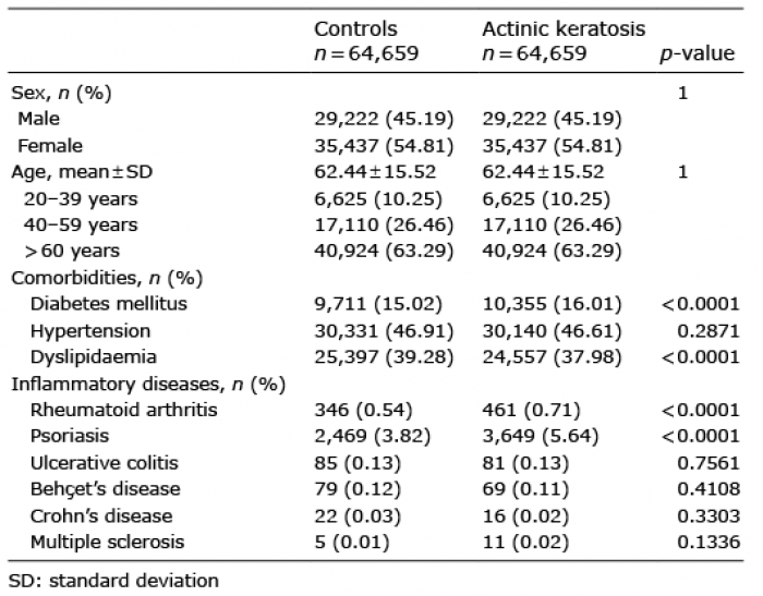
A 74-year-old man with a history of atherothrombotic brain infarction and internal carotid artery stenosis presented with intermittent claudication and leg pain. He was diagnosed with artery stenoses, and stents were placed in his left iliac artery and left superficial femoral artery. Six months later, left iliac artery stenosis was found again and percutaneous balloon angioplasty was performed (Table I). Two years after these endovascular procedures, a painful purple skin lesion appeared suddenly on his left toes (Fig. 1). No inflammatory signs, including redness and heat, were observed. Laboratory findings, including white blood cell count (WBC: 5,180/μl), eosinophils (2.1%), renal, and liver function tests were normal and C-reactive protein (CRP: 1.41 mg/dl) levels were slightly elevated. Angiographic findings had not changed compared with those reported in the previous year. He was referred to our department, and we suspected blue toe syndrome. We then performed a skin biopsy. Histopathological findings showed a mid-dermal vessel completely occluded with basophilic and convolutional foreign body gyrus-like material (Fig. 2). Although mild infiltrated lymphocytes were detected in the dermis, there were no obvious inflammatory findings, such as neutrophilic reactions, abscesses and granuloma formation. There were no other associated findings. Based on these results, the patient was diagnosed with cutaneous HPE. His skin lesions were treated with intravenous alprostadil alfadex for 1.5 months and topical sulfadiazine silver for 2 months, leading to complete recovery after 8 months. Recurrent skin symptoms were not observed for 5 months after completion of the therapies.
Table I. Details of endovascular treatment (EVT) and angiography
Fig. 1. Clinical findings. (a–c) Painful purple skin lesions on the patient’s left 2–4 toes. Brown macular and punctate purpura on the left dorsum of the foot.
Fig. 2. Histopathological findings. (a) Basophilic and convolutional foreign body materials in mid-dermal blood vessels. (Haematoxylin-eosin staining: original magnification: ×40). (b) Foreign body material manifesting as gyrus-like lesions.
HPE is induced by intravascular approaches, when the hydrophilic coatings on devices, such as endovascular indwelling catheters and stents, are stripped off as they hit vessel walls (1, 2). Polymer materials can travel to distant organs and cause occlusion, ultimately leading to sudden death; as seen in our case, cutaneous embolism alone also occur. Hydrophilic polymers are widely used as coatings for cardiac catheterization and coronary angiography medical devices, and are useful for reducing friction and preventing vasospasm (2, 4). Although hydrophilic polymers are gradually absorbed within blood vessels, the speed of absorption depends on the type of hydrophilic polymers.
A patient with HPE can be diagnosed by detecting hydrophilic polymer, present as a gyrus-like foreign body material, in blood vessels. Patients with a cholesterol embolism show purpura and cutaneous necrosis; biconvex, needle-shaped clefts are also observed as foreign-body materials (5). In this respect, clinical findings of cutaneous HPE and cholesterol embolism are similar. Although the CRP level was slightly elevated in the current case, WBC count, including eosinophil count, was normal. Laboratory data and histological findings indicated HPE rather than a cholesterol embolism. Patients with a cholesterol embolism are usually treated with oral steroids (5). In contrast, a previous report on 8 patients with cutaneous HPE showed that the cutaneous lesions resolved spontaneously without specific treatment (3). Nevertheless, if the hydrophilic polymer remains in the blood vessels beyond the acute phase it may cause an inflammatory reaction and damage vital organs. In several cases, steroids and immunomodulatory therapies have been shown to be effective in mitigating inflammatory reactions (6). In the current case, no inflammatory signs were observed, and steroid therapy was not needed. Therefore, the lack of inflammatory signs may be an indicator of spontaneous resolution or the lack of inflammatory signs may be the requirement of nonaggressive therapy. The current case demonstrates that cutaneous HPE can occur long after endovascular procedures; cases with a late-onset cholesterol embolism have also been reported (7). Therefore, distinguishing a cutaneous HPE from cholesterol embolism based on clinical findings is difficult. If we consider patients with a cholesterol embolism, skin biopsy should be performed to distinguish from cutaneous HPE. A previous study also suggests that recurrent inflammation may occur (6), and careful follow-up is therefore required.
The current patient had late-onset HPE. Most cases in previous reports of cutaneous HPE occurred within a few hours to 2 weeks (3, 8), while several cases with cutaneous HPE were observed 4–6 months after endovascular procedures (9, 10). Moreover, a case without symptoms unexpectedly presented with HPE within the small vessels 16 months after endovascular procedures on autopsy (1). A recent retrospective study demonstrated that, of 136 endovascularly examined autopsy cases, 18 (13%) showed histological evidence of HPE; however, the details of the period from the endovascular procedure to the onset and the history of endovascular procedures were not mentioned (11). Although similar studies analysing the presence of HPE in autopsy cases have been reported, HPE was detected mainly in major organs, including the lungs, and these studies did not focus on the skin in autopsy cases (11, 12). Therefore, the mechanisms of late-onset cutaneous HPE have not been clarified; however, we consider that the hydrophilic polymer would have already been present asymptomatically in the blood vessels for a long time, and subsequently travelled to the small vessels, gradually causing vascular stenosis during absorption of the hydrophilic polymer.
In conclusion, we report here a rare case of late-onset cutaneous HPE. Diagnosis of cutaneous HPE is important to determine appropriate treatment. The possibility of cutaneous HPE should be considered in patients who present skin necrosis or purpura after endovascular procedures, regardless of the onset period after the procedures.
The authors have no conflicts of interest to declare.


 Click to show fullsize
 Click to show fullsize
 Click to show fullsize