


1Noguchi Dermatology Clinic, 694-1 Uejima, Kashima-machi, Kamimashiki-gun, Kumamoto 861-3106, 2Ochanomizu Institute for Medical Mycology and Allergology, 3Department of Dermatology and Allergology, Juntendo University Graduate School of Medicine, Tokyo, 4Department of Dermatology and Plastic Surgery, Faculty of Life Sciences, Kumamoto University, 5Department of Dermatology, Japan Community Health Care Organization Kumamoto General Hospital, Kumamoto, 6Division of Bio-resources, Medical Mycology Research Center, Chiba University, Chiba and 7Department of Veterinary Dermatology, Nihon University College of Bioresource Sciences, Kanagawa, Japan. E-mail: derma@nogcli.jp
Accepted Aug 11, 2021; Epub ahead of print xx
Acta Derm Venereol 2021; 101: adv00563.
doi: 10.2340/00015555-3926
Terbinafine, which targets squalene epoxidase (SQLE), has been used to treat dermatophyte infections for approximately 30 years. In 2017, a Swiss study reported that 1% (16/1,644) of Trichophyton rubrum and 0.2% (1/412) of T. interdigitale were resistant to terbinafine (1). In 2019, we presented the first Japanese case of tinea unguium caused by a terbinafine-resistant T. rubrum isolate (Phe397Leu substitution), which was deposited as IFM 65760 (2). Our clinic obtained 3 terbinafine-resistant T. rubrum strains (Leu393Phe substitution) from 95 dermatophyte clinical isolates including T. rubrum (n = 62) and T. interdigitale (n = 33) in June 2020 (3). One strain (T. rubrum N79) was derived from a group home for individuals with intellectual disabilities. In this study, we examined the residents of this facility using mycological and molecular techniques to detect terbinafine-resistant T. rubrum strains.
The group home accommodates 54 individuals over 18 years of age and provides 24/7 care including bathing, toileting and meals. The facility is in Kumamoto, which has a subtropical climate (Köppen climate classification Cfa) with hot, humid summer at 32° north latitude. All patients diagnosed with dermatophytosis during a 6-month period (June to November 2020) were included in this study (n = 30 (20 males, 10 females); mean age 54.1 ± 17.7 years). Pathogens were identified in cultures on Sabouraud agar with chloramphenicol and cycloheximide (Mycosel agar; Kyokuto Pharmaceutical Industrial Co. Ltd, Tokyo, Japan) and/or by DNA-based detection for culture-negative onychomycosis in our clinic and Kahotechno Co., Ltd (Fukuoka, Japan). Molecular identifications, sequence analyses and antifungal susceptibility tests were performed at the Department of Veterinary Dermatology, Nihon University College of Bioresource Sciences (Kanagawa, Japan). The homology of the internal transcribed spacer region sequences in the rRNA gene of the strains was 100% (688/688 bp) identical to that of the T. rubrum reference strain IFM 63288 (GenBank, LC317851). The mutation hotspot of SQLE was determined based on the conserved sequence of T. rubrum SQLE (GenBank accession number XM_003233797) (3). The following primers were used: SQEL397S (5′- GTTGACTGGTGGCGGTATG; position 1002–1020) and SQEL397R (5′- GCTACGGAGTAAAAATGCCG; position 1315–1334) (Japanese patent application number 2021–2373). The antifungal susceptibility of the isolates to terbinafine, itraconazole, ravuconazole and luliconazole was evaluated by a broth microdilution assay according to the Clinical and Laboratory Standards Institute (CLSI) M38-A2 guidelines, with some modification (4).
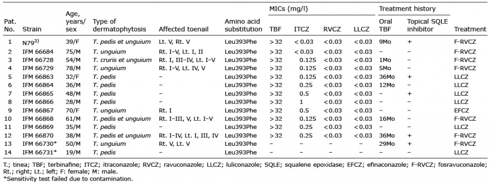
The 30 patients with dermatophytosis included patients with tinea pedis (n = 10), tinea unguium (n = 12), tinea pedis et unguium (n = 6), and tinea cruris et unguium (n = 2). Fungal cultures were positive in 17 cases and DNA-based detection of culture-negative onychomycosis was positive in 6 cases. The causative fungi were T. rubrum (n = 22, 95.6%) and T. interdigitale (n = 1, 4.3%). Among these cases, 47% (14/30) were caused by terbinafine-resistant T. rubrum. In addition, 60% (8/14) of the patients with terbinafine-resistant dermatophyte had tinea unguium and 5.4 ± 3.2 nails were affected. Regarding the affected nails, 62.5% (n = 10), 43.8% (n = 7), 50% (n = 8), 50% (n = 8), and 62.5% (n = 10) of the lesions involved the big, 2nd, 3rd, 4th and 5th toenail, respectively. Most patients (8/14) with terbinafine-resistant dermatophytes received oral terbinafine (125 mg, daily) for a mean duration of 24.4 ± 14.3 months. Five patients (5/14) were treated with topical SQLE inhibitors, either alone or in combination with oral treatment. Topical regimens included terbinafine, bifonazole, butenafine and liranaftate. Five patients (5/14) had no history of oral terbinafine or topical SQLE inhibitor use. The characteristics of patients with terbinafine-resistant isolates are shown in Table I. Nucleotide substitution within SQLE (1177TTA→TTC) was consequently detected in all 14 terbinafine-resistant strains, leading to Leu393Phe substitution in T. rubrum SQLE proteins. These isolates were deposited in Chiba University with IFM numbers. The minimum inhibitory concentrations (MICs) for the mutant strains were > 32 mg/l for terbinafine, < 0.03–1 mg/l for itraconazole, < 0.03 mg/l for luliconazole and < 0.03 mg/l for ravuconazole.
Table I. Characteristics of patients with terbinafine-resistant Trichophyton rubrum isolates
Terbinafine is the most commonly prescribed oral antifungal medicine that is approved for the treatment of tinea unguium. Meanwhile, terbinafine-resistant tinea corporis due to prolonged terbinafine therapy has been reported in a 62-year-old man with Darier disease (5). In our study, cases 5 and 12 received oral terbinafine for 3 years. In particular, case 12, who had hyperkeratotic-type tinea pedis and severe tinea unguium, might have been an index patient. Because Japanese people remove their shoes and walk barefoot in tatami rooms, the infection may spread even in a living room (6).
Itraconazole resistance in T. rubrum depends not on mutation of the target enzyme (lanoconazole 14-α-demethylase), but on the overexpression of the TruNDR2 gene, which encodes multidrug transporters of the ABC family (7). Itraconazole-resistant T. rubrum (TIMM20092) was isolated from a tinea pedis patient in Switzerland with a minimum inhibitory concentration (MIC) of 0.5 mg/l for itraconazole (7, 8). In our study, the MIC for itraconazole ranged from
Fosravuconazole, which was approved for tinea unguium treatment by the Japanese government in 2018 (9), is a novel triazole antifungal drug developed as a water-soluble prodrug for ravuconazole (10). Its excellent oral absorbability and systemic bioavailability have resulted in high serum drug concentrations and a long half-life (10). We treated 7 severe cases of tinea unguium with oral fosravuconazole and 1 mild case with efinaconazole 10% solution (11). Six patients with tinea pedis were treated with luliconazole 1% cream. As of July 2021, among the 7 fosravuconazole-treated patients with tinea unguium, 3 had been successfully cured, while 2 were showing a positive improvement. In addition, one patient treated with efinaconazole was also found to be improving. Among the 6 luliconazole-treated patients with tinea pedis, 5 had been successfully cured, while the other patient with a hyperkeratotic type had begun a treatment regimen with oral fosravuconazole.
A questionnaire survey of antifungal-resistant dermatophytes in representatives from European countries revealed that 85% of all countries (17/20) observed clinical and/or mycological confirmed resistance to terbinafine in 64% (61/96) and to itraconazole in 41% (39/96), while also observing resistance to fluconazole in 16% (15/96) (12). The 3 prevalent species were T. rubrum (33/95), Microsporum canis (23/95), and T. mentagrophytes (17/95) (12). Dermatologists should focus on the increasing numbers of terbinafine-resistant dermatophytes.
This work was partly supported by the Japan Agency for Medical Research and Development, AMED under Grant Number JP21fk0108094. The authors thank Dr. Yoshiharu Ohsato (Kahotechno Co., Ltd., Fukuoka, Japan) for the genetic diagnosis of onychomycosis.


 Click to show fullsize