


Andrea Giuliani1, Gaspare Galati1, Leoluca Parisi2, Teresa Ricciardulli1, Michelangelo Bartolo2, Elvira Tartaglia1, Miriam Grimaldi3 and Guglielmo Pranteda2*
University of Rome ‘‘La Sapienza’’, 1I School of Medicine, Department of Surgery ‘‘Pietro Valdoni’’, 2Department of Neurology and ORL, Umberto I Polyclinic, and 3II School of Medicine, Department of Skin and Sexually-transmitted Diseases and Plastic and Reconstructive Surgery, S. Andrea Hospital, Via di Grottarossa 1035, Rome, Italy. *E-mail: cpranteda@hotmail.com
Accepted May 6, 2005.
Sir,
Herpes zoster (HZ) is caused by reactivation of latent infection with varicella zoster virus established in the sensory ganglia after an earlier primary infection with chickenpox. The usual neurological postherpetic complication is a dermatomal distribution pain that persists for months to years after resolution of HZ rash. Although segmental motor paralysis, both visceral and somatic, is an unusual and less well known complication, it sometimes occurs in the same or in the adjacent involved dermatomes (1). We present a case of HZ complicated by motor involvement of T10–T11 segments with an abdominal bulge mimicking a herniation.
CASE REPORT
A 75-year-old man presented a left flank wall herniation that had gradually developed in size over the previous 2 months and burning pain over the involved area. Four months before the consultation he suffered from left flank HZ rash complicated by persistent neuralgia. At that time, therapy with oral acyclovir was administered (800 mg five times daily for 1 week) with regression of the skin eruption. Five years before, he was curatively resected for a sigmoid colon cancer through a midline laparotomy and subsequently received cycles of systemic chemotherapy.
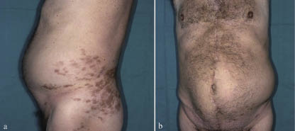
On physical examination, the skin evidenced hyperpigmentated macules in the dermatomes previously involved by herpetic rash; a reducible, painful swelling, 20 cm × 15 cm in diameter, increasing in prominence with intra-abdominal straining, protruded in an area of flaccid muscles of the left flank (Fig. 1). The laparotomy incision evidenced uncomplicated healing. General laboratory tests and the tumour markers were normal. Thoraco-abdominal computed tomography (CT) showed no abdominal mass, the flank wall bulged asymmetrically and the left abdominal large muscle was thinner than the right side. Thoracic and lumbar spine magnetic resonance imaging (MRI) evidenced no mechanical nerve root damage. On neurological examination superficial abdominal reflexes were absent in the left side and hyperaesthesia was present in this area. The sensory examination was otherwise normal. Concentric needle electromyography (EMG) of external and internal oblique abdominal muscles, rectus abdominis muscles and paravertebral thoracic muscles from T9 to L1 revealed membrane irritability with positive waves and fibrillations of left-sided muscles, most prominently at T10–T11 level. Motor unit action potentials were present in left-sided muscles, although reduced in number and in amplitude. EMG recordings on the right side were normal. The neurological findings were consistent with an involvement of left anterior roots at T10–T11 level by shingles. A treatment with intravenous betametasone was started at a dosage of 4 mg daily for 10 days, then 1.5 mg daily for a further 5 days, with partial improvement of the pain. On a follow-up examination, 7 months after the onset, the bulge was partially resolved and the patient reported a significant resolution of the pain and hyperaesthesia.
Fig. 1. Lateral view showing postherpetic hyperpigmented maculas of the left flank (a) and frontal view showing unilateral abdominal swelling (b).
DISCUSSION
Although HZ usually affects sensory nerves, motor deficit during the infection is reported in 5% of cases (1). This percentage increases in the elderly and in immunocompromised patients, and it evidences a correlation with the dermatomes involved (2). The trunk is a major area of involvement with HZ; however, the postherpetic motor complication of thoraco-abdominal muscles is considered a rare event. An electromyographic evaluation, however, evidenced it in 35% of thoracic HZ cases (3). Visceral motor complications are reported, particularly neurogenic bladder dysfunction and colonic pseudo-obstruction (4). The postherpetic paresis of abdominal wall muscles has a prevalence estimated to be around 0.17% of cases (5) and it presents as an abdominal or flank bulge (6–10). As regards our patient, the differential diagnoses were a surgical nerve injury or a neural involvement by a recurrence of previous colon disease. The time elapsed from the surgery to the occurrence of swelling and the type of the surgery performed for the colon cancer excluded a surgical nerve injury. MRI did not show nerve root compression. After a 7-month follow-up, our patient had partial regression of the bulge and a significant improvement of neurological symptoms. Prognosis of postherpetic motor paralysis seems to be good, with a complete or partial spontaneous recovery in 50–70% of patients within 1 year of the onset (4). In our experience EMG and CT were helpful to explain the aetiopathogenesis of abdominal bulge mostly because a muscle herniation was possible in a patient with previous laparotomy.
REFERENCES
1. Thomas JE, Howard FM. Segmental zoster paresis – a disease profile. Neurology 1972; 22: 459–466.
2. Devinsky O, Cho ES, Pelito CK, Price RW. Herpes zoster myelitis. Brain 1991; 114: 1181–1196.
3. Cioni R, Giannini F, Passero S, Paradiso C, Rossi S, Fimiani M, et al. An electromyographic evaluation of motor complications in thoracic herpes zoster. Electromyogr Clin Neurophysiol 1994; 34: 125–128.
4. Akiyama N. Herpes zoster infection complicated by motor paralysis. J Dermatol 2000; 27: 252–257.
5. Barroso FA. Abdominal distension due to herpes zoster. Medicina (B Aires) 2002; 62: 53–54.
6. Tjandra J, Mansel RE. Segmental abdominal herpes zoster paresis. Aust N Z J Surg 1986; 56: 807–808.
7. Jandolo B, Biolcati G, Montanari U, Pietrangeli A, Fazio M. Segmental abdominal zoster paresis. Riv Neurol 1987; 57: 274–276.
8. Gottschau P, Trojaborg W. Abdominal muscle paralysis associated with herpes zoster. Acta Neurol Scand 1991; 84: 344–347.
9. Vincent KD, Davis LS. Unilateral abdominal distension following herpes zoster outbreak. Arch Dermatol 1998; 134: 1168–1169.
10. Molinero J, Nagore E, Obon L, Miquel FJ, Aliaga A. Metameric motor paresis following abdominal herpes zoster. Cutis 2002; 69: 143–144.

