Başak Yalcçın, Emine Tamer, Güneş Gür, Pınar Öztas, Muhterem Üstün Polat and Nuran Allı
Ankara Numune Education and Research Hospital 1st Dermatology Clinic, Kehribar sok. Mesa Yamac¸ 1 sit., No: 11/15 Gaziosmanpaşa, TR-06700, Ankara, Turkey. E-mail: yalcinbasak@yahoo.com
Accepted June 7, 2005.
Sir,
Neurofibromatosis 1 (NF1) is an autosomal dominant disease which predominantly involves the skin and the nervous system. The cardinal features of NF1 include neurofibromas, café-au-lait spots, axillary and inguinal freckling, eye abnormalities comprising Lisch nodules, optic glioma and osseous lesions and learning disabilities (1). Noonan syndrome (NS), is a genetic disorder whose prevelance is estimated to be 1 in 1000 or 2500 live births. It is characterized by unusual triangular-shaped face, hypertelorism, down-slanting eyes, ptosis, strabismus, amblyopia, refractive errors, low-set ears with thickened helices, high nasal bridge, webbed neck, congenital heart disease (dysplastic/stenotic pulmonic valve, hypertrophic cardiomyopathy), short stature and chest deformities (pectus carinatum/excavatum, scoliosis) (2). Vitiligo, characterized by depigmented macules and patches on the skin, has been commonly reported as a component of multiple autoimmune syndromes and in association with autoimmune thyroid disease such as Hashimoto’s thyroiditis and Graves’ disease (3). Although NF1 has been seen in relation with various autoimmune diseases, coexistence with either Hashimoto’s disease or vitiligo has not been reported previously. Here we report a case with established NF1/NS who also has a diagnosis of vitiligo and Hashimoto’s disease.
CASE REPORT
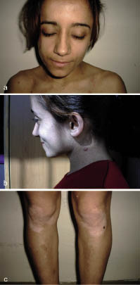
A 20-year-old female patient was admitted to the hospital with hyperpigmented skin patches that had been present since birth and tumoral formations on her skin that had recently increased in number. She had also developed hypopigmented patches on her knees and elbows 4 years previously. Her family history revealed that her mother and father were second degree relatives (maternal cousins) and one of her cousins had similar skin lesions. On her physical examination she was 145 cm tall. She had scoliosis in her thoracic vertebrae and a short webbed neck. Her face was triangular with a prominent forehead and a small chin (Fig. 1a). Her ears were small and low-set with thickened helices (Fig. 1b). Her eyes were down-slanting and ptotic. Her dermatological examination revealed bilateral axillary freckling, several hyperpigmented macules and patches between 1 and 5 cm in diameter which were consistent with café-au-lait spots. She had large patches of depigmentation between 5 and 10 cm in diameter on the knees and elbows consistent with vitiligo (Fig. 1c). Skin biopsy confirmed the diagnosis of vitiligo. On laboratory examination, karyotype analyses revealed normal female with 46 XX. Complete blood count, serum and urine biochemistry were within normal limits. Thyroid stimulating hormone was 2.06 µIU/ml (normal range 0.35–4.95); T3 was 172 ng/dl (normal range 95–190); and T4 was 7.54 µg/dl (normal range 5–11). Antithyroid peroxidase level was 528 IU/ml (normal range 0–100 IU/ml) and antithyroglobulin was 442 IU/ml (normal range 0–100 IU/ml). The thyroid gland was firm on palpation and ultrasonography revealed a diffuse mildly enlarged gland. Abdominopelvic ultrasonography was normal. Cranial magnetic resonance imaging (MRI) disclosed hyperintense non-specific signal abnormalities in right posterior paraventricular white matter in T2-weighted sequences. Electrocardiographic and echocardiographic examinations were normal.
Fig. 1. (a) Atypical triangular face with prominent forehead and small chin, ptotic and down-slanting eyes, webbed neck, café-au-lait spots and neurofibromas. (b) Small low-set ear with thickened helix. (c) Depigmented vitiligo patches on knees. (Reproduced with the patient’s permission.)
DISCUSSION
The coexistence of NF1 and NS has been reported previously but the true incidence remains unknown (4, 5). It also remains unclear whether NF1/NS represents a form of NF1 (with mutations in the NF1 gene) or is a separate syndrome (6). In NS, cardiovascular system involvement is present in >50% of patients. The primary source of morbidity and mortality in these patients depends on the presence and type of congenital heart disease (2). However, cardiovascular involvement and congenital heart diseases are not expected in patients with NF1. We did not detect a cardiovascular pathology either clinically or by laboratory examinations in our patient. The NF1 gene is located on chromosome 17q11.2. Neurofibromin, the product of the non-mutated gene, has the activity of a GTPase-activating protein and is capable of down-regulating the cellular p21-ras proto-oncogene. The loss of neurofibromin function may lead to uncontrolled cell growth or tumour formation, which may lead to increased tumour formation in patients with NF1, including both benign and malignant peripheral nerve sheath tumours and some extracutaneous tumours such as phaeochromocytoma, carcinoid syndrome, rhabdomyosarcoma, juvenile chronic myelogeneous leukaemia and lung cancers (7, 8). Furthermore, abnormal neurofibromin production suppresses fas-ligand expression. This may prevent apoptosis of CD4+ T cells, which is important in the development of autoimmunity (9). Our case had two coexisting autoimmune diseases – vitiligo and Hashimoto’s disease. We have not encountered a similar association in the literature. However, some other autoimmune diseases such as Graves’ disease, autoimmune haemolytic anaemia, myasthenia gravis, systemic lupus erythematosus and bullous pemphigoid were reported in patients with NF1 (10–14). The coexistence of Hashimoto’s disease and vitiligo in our patient may be a coincidence.
However, decreased T-cell apoptosis due to abnormal neurofibromin production may be an underlying factor for the development of the autoimmune diseases in our patient. As the number of reports on the coexistence of NF1 and autoimmune diseases increases, an association rather than a coincidence becomes more likely.
REFERENCES
1. Pivnick EK, Riccardi VM. The neurofibromatoses. In: Freedberg IM, Eisen AZ, Wolff K, Austen KF, Goldsmith LA, Katz SI, et al., eds. Dermatology in general medicine. New York: McGraw-Hill, 1999: 2152–2158.
2. Happle R. Neurocutaneous diseases. In: Freedberg IM, Eisen AZ, Wolff K, Austen KF, Goldsmith LA, Katz SI, et al., eds. Dermatology in general medicine. New York: McGraw-Hill, 1999: 2131–2148.
3. Betterle C, Caretto A, De Zio A, Pedini B, Veller- Fornasa C, Cecchetto A, et al. Incidence and significance of organ specific autoimmune disorders (clinical, latent or only autoantibodies) in patients with vitiligo. Dermatologica 1985; 171: 419–423.
4. Allanson JE, Hall JG, Van Allen MI. Noonan phenotype associated with neurofibromatosis. Am J Med Genet 1985; 21: 457–462.
5. Colley A, Donnai D, Evans DG. Neurofibromatosis/Noonan phenotype: variable features of type 1 neurofibromatosis. Clin Genet 1996; 49: 59–64.
6. Baralle D, Mattocks C, Kalidas K, Elmslie F, Whittaker J, Lees M, et al. Different mutations in the NF1 gene are associated with neurofibromatosis-Noonan syndrome (NFNS). Am J Genet A 2005; 119: 1–8.
7. Schwarz J, Belzberg AJ. Malignant peripheral nerve sheath tumors in the setting of segmental neurofibromatosis. Case report. J Neurosurg 2000; 93: 530–532.
8. Yalcin B, Toy GG, Tamer E, Oztas P, Koc D, Dikicier B, et al. Acceleration of segmental neurofibromatosis in bronchoalveolar lung carcinoma. Dermatology 2004; 209: 342.
9. Abbas A, Lichtman AH. Disease caused by immune responses: hypersensitivity and autoimmunity. In: Abbas A, Lichtman AH, eds. Cellular and molecular immunology, 5th edn. Philadelphia: Saunders, 2005: 411–431.
10. Bolko P, Wasko R, Waligorska J, Sowioski J. Graves’ disease and hyperprolactinemia in a patient with Noonan syndrome neurofibromatosis type 1. Ann Endocrinol (Paris) 2004; 65: 121–124.
11. Tekin F, Ozutemiz O, Carcurgan S, Ilter T. Autoimmune haemolysis as an unusual cause of anaemia in von Recklinghausen’s disease. Neth J Med 2004; 62: 337–339.
12. Ferner RE, Hughes RA, Johnson MR. Neurofibromatosis 1 and multiple sclerosis. J Neurol Neurosurg Psychiatry 1995; 58: 582–585.
13. Corominas H, Guardiola JM, Matas L, Vazquez G. Neurofibromatosis and systemic lupus erythematosus. A matter of coincidence? Clin Rheumatol 2003; 22: 496–497.
14. Yesudian PD, Wilson NJ, Parslew R. Bullous pemphigoid and neurofibromatosis – a chance association requiring special vigilance. Clin Exp Dermatol 2000; 25: 658–659.