


Maria Esposito, Elisabetta Capriotti, Alessandro Giunta, Luca Bianchi and Sergio Chimenti*
Department of Dermatology, University of Roma ‘‘Tor Vergata’’, Viale Oxford 81, IT - 00133, Rome, Italy. *E-mail: chimenti@dermatologica.it
Accepted June 20, 2005.
Sir,
Although the systemic administration of corticosteroids (CCS) and immunosuppressive agents has dramatically improved the prognosis of pemphigus vulgaris (PV), the disease is still associated with a significant morbidity and mortality (1, 2). Rituximab is a genetically engineered chimeric murine anti-CD20 monoclonal antibody (mAb) that targets B cells. This effect leads to a strong reduction in antibody production and has shown efficacy in B-cell lymphomas, plasma cell-dependent diseases and some autoimmune disorders (3). Herein we describe two patients affected by PV who were unresponsive to standard therapies treated with rituximab.
CASE REPORTS
Patient 1. In September 2003, a 45-year-old man was first admitted for a severe flare of PV. Results of clinical examination revealed extensive blisters and crusted erosions primarily involving the scalp, the face, the trunk and the upper extremities (Fig. 1a). Diagnosis was confirmed by histology and direct and indirect immunofluorescence (IF) microscopy studies. The patient serum was previously tested by a commercially available enzyme immunoassay (EIA), based on recombinant desmogleins (Dsg) and the presence of antibodies to Dsg-1 (index: 128, normal values: 0–14) and Dsg-3 (index: 155, normal values: 0–7) was demonstrated. Disease severity was estimated as severe, with a score of 8 using the Herbst & Bystryn disease severity grading system, ranging from 0 to 10 (4). The patient had a history of PV since 1993 with poor clinical benefits from treatments and very short disease-free periods. Previous therapies included low-dose prednisone in monotherapy or in combination with azathioprine, followed by cyclosporin A and mycophenolate mofetil. The disease rapidly progressed and the patient was switched to methotrexate in association with intravenous immunoglobulin (IVIG) infusions, but the beneficial effects were short-lived and IVIG infusions were interrupted. After a 3-month period of maintenance therapy with methotrexate followed by a further 2 weeks without therapy, the first intravenous treatment with rituximab was administered at 375 mg/m2 body surface area, and repeated weekly four times. Before each infusion, the patient was premedicated with intravenous chlorpheniramine maleate at a dose of 20 mg, to prevent or minimize reactions to the chimeric mAb. No other drugs were given to the patient to further increase the clinical efficacy during the course of treatment (1 month) and the follow-up (7 months). After the second infusion of rituximab (week 2), lesions gradually healed, reaching complete resolution after 4 weeks on the trunk and upper extremities and partial remission on the scalp. New blisters have not developed so far. The clinical condition of the patient continued to improve over the next 7 months (Fig. 1b). At 7 months of follow-up, only one small lesion was still present on the forehead.
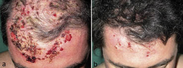
Fig. 1. Patient 1 at baseline (a) showing crusted erosions and blisters involving the scalp and the forehead. After 5 months of follow-up (b) the scalp and forehead show an almost complete healing of lesions associated with a significant regrowth of hair.
After the second infusion, B-cell levels, monitored monthly in peripheral blood, became undetectable, remaining so for up to 7 months. An assay with primate oesophagus as substrate did not detect any significant change of circulating anticytoplasmic membrane autoantibodies (Nova Lite™ SA. A. Menarini Diagnostics), which remained between 1:160 and 1:80 over the follow-up period. At 7 months of follow-up the EIA index of anti-Dsg-1 was 80, while anti-Dsg 3 was 92.
Patient 2. A 49-year-old man was referred to our department in April 2003 because of a 2-year history of severe PV. He presented with widespread mucocutaneous disease with diffuse blisters, erosions and crusting of the scalp and the upper trunk and severe painful mucous membrane erosions of the oral cavity which caused dysphagia and weight loss. The diagnosis of PV was confirmed by histology and direct and indirect IF microscopy studies. The patient had circulating autoantibodies against the epithelial cell surface of primate oesophagus at a titre of 1:320. The patient had severe disease with a severity score of 7 (4). The patient had been previously treated unsatisfactorily with an increasing regimen of systemic corticosteroids, azathioprine and cyclosporin A. Subsequently, the patient was treated with IVIG, but he experienced side effects which led to interruption of the treatment. Therefore, after 3 months of wash-out period, four rituximab infusions were given intravenously at a dose of 375 mg/m2 body surface on a weekly basis. No other drug administration was required during the treatment and the following 6 months. A clinical improvement of the skin lesions located on the scalp and trunk was observed from the third week of treatment (score value from 4 to 3 at week 4, 1 at week 16, 0 at week 24). The response was considered maximal after 4 months of follow-up with a complete healing of the skin lesions, without new blister development. The mucosal involvement has shown a complete resolution during the follow-up period. As in patient 1, after the second infusion, circulating B cells became undetectable and remained so for 6 months. The titre of circulating antibodies against the epithelial cell surface of primate oesophagus showed no significant change, as it varied from 1:320 to 1:160 during the follow-up.
DISCUSSION
PV is due to B-cell production of autoantibodies directed primarily against Dsg-3 and Dsg-1 (5, 6). Until the introduction of systemic corticosteroids, PV was an almost invariably fatal disease with a mortality rate up to 90% (1, 2). Immunosuppressive treatments have resulted in a dramatic improvement in prognosis (2). However, the combination of drugs is frequently associated with side effects (2). Furthermore, in some cases these agents are ineffective or contraindicated.
Rituximab is a chimeric mAb with human IgG1 constant regions and murine light and heavy chain variable regions. It has shown a high efficacy in the treatment of relapsing and refractory follicular and high grade lymphomas (3, 7). The antibody targets the B-cell transmembrane antigen CD20, located on pre-B, mature B lymphocytes and most B-cell neoplasms, and is able to block the disease process by eliminating CD20 B cells from peripheral blood, provoking their lysis via both complement-dependent and antibody-dependent cellular cytotoxicity (3, 7). Studies on PV have shown that patients with severe disease often have high levels of anti-Dsg3 IgG-producing B cells in the peripheral blood (6), which may explain the therapeutic effect of rituximab (8). Analogously treatment of paraneoplastic pemphigus associated with follicular B-cell lymphoma has been described (9, 10). Subsequently, few cases of severe and refractory PV have been reported with satisfactory results and a tolerable adverse effect profile (8, 11–13, 15, 16) (Table I). In the clinical management of PV, antibody titres can be used for monitoring therapeutic efficacy because they often correlate with disease activity (14). However, despite the early clinical benefits and drastic peripheral B-cell depletion, as seen in our patients, circulating autoantibodies against the epithelial cell surface of primate oesophagus used as substrate did not significantly decrease. Speculatively the coexistence of clinical remission with persisting low antibody titre may be explained, assuming that autoantibodies require the presence of B cells to extend their effect (6, 8). Cutaneous complete remission still persist in both patients, 6 and 7 months after treatment; serious adverse effects, especially infections, were not observed during the treatment and the follow-up period.
As regards the long-term efficacy of rituximab in the treatment of PV, only two cases have been reported so far, while tapering previous therapies (15, 16). As far as we know our experience is the first report of a long-term remission of PV after treatment with rituximab as monotherapy.
Table I. Published cases of pemphigus vulgaris treated with rituximab
Length of
Ref. no. Patient Age Infusions Regimen of administration Clinical result Follow-up efficacy
8 F 29 6 Polychemotherapy (CCS, MMF, CP) Almost complete NA NA
11 M 54 4 Polychemotherapy (MMF, CCS) Almost complete NA NA
12 F 54 4 Polychemotherapy, (CCS) Good clinical improvement NA NA
15 F 53 4 Polychemotherapy (CCS, CP) Complete remission 40 weeks 40 weeks
13 F 34 8 Polychemotherapy (CCS) Significant improvement 36 weeks 16 weeks
F 42 8 Polychemotherapy (CCS, CsA) Complete remission 24 weeks 24 weeks
M 20 4 Polychemotherapy (CCS) Significant improvement 24 weeks 24 weeks
16 M 39 4 Polychemotherapy (PE, CCS) Complete remission 40 weeks 40 weeks
This M 45 4 Monotherapy Almost complete remission 28 weeks 28 weeks report M 49 4 Monotherapy Complete remission 24 weeks 24 weeks
M, male; F, female; NA, no data available; CCS, corticosteroids; MMF, mycophenolate mofetil; CP, cyclophosphamide; CsA, cyclosporin A; PE, plasma exchange.
REFERENCES
1. Lever WF. Pemphigus and pemphigoid. Springfield, IL: Charles C Thomas, 1965.
2. Fine JD. Management of acquired bullous skin disease. N Engl J Med 1995; 333: 1475–1484.
3. Johnson PW, Glennie MJ. Rituximab: mechanism and applications. Br J Cancer 2001; 85: 1619–1623.
4. Herbst A, Bystryn JC. Pattern of remission in pemphigus vulgaris. J Am Acad Dermatol 2000; 42: 422–427.
5. Amagai M, Karpati S, Prussick R, Klaus-Kovtun V, Stanley JR. Autoantibodies against the aminoterminal cadherin-like binding domain of pemphigus vulgaris antigen are pathogenic. J Clin Invest 1992; 90: 919–926.
6. Kitajima Y. Current and prospective understanding of clinical classification, pathomechanism and treatment in pemphigus. Arch Dermatol Res 2003; 295: S17–S23.
7. Coiffier B, Haioun C, Ketterer N, Engert A, Tilly H, Ma D. Rituximab (anti-CD20 monoclonal antibody) for the treatment of patients with relapsing or refractory aggressive lymphoma: a multicenter phase II study. Blood 1998; 92: 1927–1932.
8. Salopek TG, Logsetty S, Tredget EE. Anti-CD20 chimeric monoclonal antibody (rituximab) for the treatment of recalcitrant, life-threatening pemphigus vulgaris with implications in the pathogenesis of the disorder. J Am Acad Dermatol 2002; 47: 785–788.
9. Heizmann M, Itin P, Wernli M, Borradori L, Bargetzi MJ. Successful treatment of paraneoplastic pemphigus in follicular NHL with rituximab: report of a case and review of treatment for paraneoplastic pemphigus in NHL and CCL. Am J Hematol 2001; 66: 142–144.
10. Borradori L, Lombardi T, Samson J, Girardet C, Saurat JH, Hugli A. Anti-CD20 monoclonal antibody (rituximab) for refractory erosive stomatitis secondary to CD20 (+) follicular lymphoma-associated paraneoplastic pemphigus. Arch Dermatol 2001; 137: 269–272.
11. Cooper HL, Healy E, Theaker JM, Friedmann PS. Treatment of resistant pemphigus vulgaris with an anti- CD20 monoclonal antibody (rituximab). Clin Exp Dermatol 2003; 28: 366–368.
12. Herrmann G, Engert A, Hunzelmann N. Treatment of pemphigus vulgaris with anti-CD20 monoclonal antibody (rituximab). Br J Dermatol 2003; 148: 602–603.
13. Dupuy A, Viguier M, Bedane C, Cordoliani F, Blaise S, Aucouturier F, et al. Treatment of refractory pemphigus vulgaris with rituximab (anti-CD20 monoclonal antibody). Arch Dermatol 2004; 140: 91–96.
14. Sams WM Jr, Jordon RE. Correlation of pemphigoid and pemphigus antibody titres with activity of disease. Br J Dermatol 1971; 84: 7–13.
15. Virgolini L, Marzocchi V. Anti-CD20 monoclonal antibody (rituximab) in the treatment of autoimmune diseases. Successful result in refractory pemphigus vulgaris: report of a case. Haematologica 2003; 88: ELT24.
16. Espana A, Fernandez-Galar M, Lloret P, Sanchez-Ibarrola A, Panizo C. Long-term complete remission of severe pemphigus vulgaris with monoclonal anti-CD20 antibody therapy and immunophenotype correlations. J Am Acad Dermatol 2004; 50: 974–976.

