


Evmorfia Ladoyanni1, Jonathan North2 and Chin Y. Tan1
1Birmingham Skin Centre and 2Department of Immunology, City Hospital, Dudley Road, Birmingham, UK. E-mail: Effil@doctors.org.uk
Accepted June 8, 2006
Sir,
Idiopathic CD4+ T-cell lymphocytopaenia (ICL) is a diagnosis of exclusion and very rare (1). Squamous cell carcinoma (SCC), Bowen’s disease and viral warts (2–4) have been reported in ICL. Here we report a patient with ICL who has developed a spectrum of human papilloma virus (HPV)-related skin problems over the last 17 years and his management.
CASE REPORT
A 49-year-old man presented in 1998 with a 10-year history of multiple recalcitrant viral warts on his hands, groin and scrotal area. He also had an SCC in situ excised from his scrotum in 1995 and from his left palm in 1998. There was no other history of note. ICL was considered. Immunological tests showed a low CD4+ count of 420 cells/mm3 (normal 700–1000 cells/mm3) equal to less than 16% of total CD count and a slightly raised CD8+ count of 930 cells/mm3 (normal 500–900 cells/mm3). Tests for human immunodeficiency virus (HIV)1 and HIV2 as well as human T-cell lymphotropic virus (HTLV)1 and HTLV2 were negative. Serum immunoglobulins were normal. The diagnosis of ICL was established. Since 2000 he has developed 3 further SCC in situ on the scrotum and left palm (Fig. 1), 3 condylomata accuminata on the suprapubic area, groin and thigh (Fig. 2), multiple intra-epithelial carcinomas on the scrotum, suprapubic area, lower abdomen and waistline and multiple viral warts on the hands and fingers (Fig. 1). His therapy so far has consisted of: imiquimod for the lesions on the scrotum, with very good response; fluoruracil cream and photodynamic
therapy to a lesion on the thigh, with moderate response;
multiple cryotherapy for viral warts on the hands and for extensive Bowen’s disease on the lower abdomen and suprapubic area; and multiple excisions, curettage and cautery. He is currently on oral acitretin 50 mg daily with moderate response. His most recent CD4+ count is 16% of the total CD count. The CD8+ count is still mildly raised.
Fig. 1. Squamous cell carcinoma in situ and multiple viral warts on the left palm.
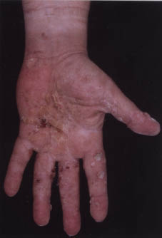
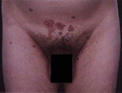
Fig. 2. Multiple lesions including condyloma accuminata in the suprapubic area.
DISCUSSION
ICL is a diagnosis of exclusion and very rare (1). It was first established in 1992 and comprises the following criteria for diagnosis: a reduced number of circulating CD4+ T-lymphocytes in more than one sample, < 300 cells/mm3 or < 20% of total T-cells; no laboratory evidence of infection with HIV1 and HIV2 nor HTLV1 or HTLV2; the absence of any other defined immunodeficiency or therapeutic management associated with depressed CD4+ T-cells. The gold standard therapy for ICL remains to be established. Symptomatic treatment and an aggressive approach to opportunistic pathogens are recommended.
SCC, Bowen’s disease and viral warts have been reported in ICL (2–6). Our patient exhibits a spectrum of recalcitrant HPV-related skin problems with no mucosal involvement. This is very rare. He has been fortunate, as so far his main problems have involved only his skin. We attribute this to the degree of immunosuppression, which is mild. The role of HPV infection as a possible aetiological agent for ICL is not widely accepted (6, 7) and recurrent warts and condylomata caused by various HPV types have been associated with poor local or systemic cellular immunosuppression. More recent studies suggest that ICL might be due to accelerated programmed T-cell death by apoptosis (8). Epidermodysplasia verruciformis (EV), a rare genodermatosis, is a genetically determined, life-long HPV infection of the skin due to a marked decrease in cell-mediated cytotoxicity against EV HPV harbouring keratinocytes (9). Skin cancers in EV show a very slow progression and are most commonly only locally destructive. Mucosal involvement is not seen. We feel the natural history of our patient can be compared to that of EV HPV. His management so far has been challenging and regular long-term follow-up is mandatory.
REFERENCES
1. Famularo G, Giacomelli R, De Simone C, Tonietti G. The syndrome of CD4+ lymphocytopenia. Ann Ital Med Int 1994; 9: 22–26.
2. Hagashi T, Hinoda Y, Takahashi T, Adachi M, Miura S, Izumi T, et al. Idiopathic CD4+ T- lymphocytopenia with Bowen’s disease. Int Med 1997; 36: 822–824.
3. Smith DK, Neal JJ, Holmberg SD and The Centres for Disease Control Idiopathic CD4+ T lymphocytopenia Task Force. Unexplained opportunistic infections and CD4+ T-lymphocytopenia without HIV infection. An investigation of cases in the United States. N Engl J Med 1993; 328: 380.
4. Hardman CM, Baker BS, Lortan J, Breuer J, Surentheran T, Powles A, Fry L. Active psoriasis and profound CD4+ lymphocytopenia. Br J Dermatol 1997; 136: 930–932.
5. Stetson CL, Rapini RP, Tyring SK, Kimborough RC. CD4+ T lymphocytopenia with disseminated HPV. J Cutan Pathol 2002; 29: 502–505
6. Manchado Lopez P, Ruiz de Morales JMG, Ruiz
Gonzalez I, Rodriguez Prietro MA. Cutaneous infections by papillomavirus, herpes zoster and Candida albicans as the only manifestation of idiopathic CD4+ T lymphocytopenia. Int J Dermatol 1999; 38: 119–121.
7. Trying KS. Cellular immune responses in human
papillomavirus infections. In: Burgdorf WH, ed. Dermatology
progress and perspectives. New York: Parthenon, 1992: p. 218–222.
8. Laurence J, Mitra D, Steiner M, Lynch DH, Siegal FP, Staiano-Coico L. Apoptotic depletion of CD4+ T Lymphocytopenia. J Clin Invest 1996; 97: 672–680.
9. Majewski S, Jablonska S, Orth G. Epidermodysplasia Verruciformis. Immunological and nonimmunological surveillance mechanisms: role in tumor progression. Clin Dermatol 1997; 15: 321–334.

