


Claire Bernier1, Jean-Michel Nguyen2, Gaëlle Quéreux1, Jean-Jacques Renault1, Brigne Bureau1 and Brigitte Dreno1
1Unit of Skin Oncology, Hôtel Dieu, Place Alexis Ricordeau, and 2Clinical Research Department, Methodological Unit, Immeuble Deurbroucq, Nantes, France
Making a differential diagnosis between early mycosis fungoides (MF) and parapsoriasis is often difficult at the clinical and histological level. The aim of this study was to explore markers that could help in this process. A total of 88 patients were included in 2 categories: large plaque parapsoriasis and digitiform parapsoriasis. A histological examination was performed for each patient, and expression of the antigen My7 (CD13), which is lacking in cutaneous T-lymphomas (CTCL) (but not in inflammatory lesions) and rearrangement of the T-cell receptor gene were analysed. A histological aspect of epidermotropic CTCL was observed in 23.5% of cases of large plaque parapsoriasis and 15% of cases of digitiform parapsoriasis. A disappearance of My7 antigen was noted in the 2 forms of parapsoriasis, more frequently when there was CTCL histology. A cutaneous clone was observed in 10.3% of cases of large plaque parapsoriasis, but not of digitiform parapsoriasis. For 3 patients, a cutaneous clone and a disappearance of My7 were associated with a non-specific histology. Considering these histological, immunological and molecular biological data, it appears that My7 antigen combined with T-cell clone may help the dermatologist to confirm the diagnosis of early MF. Moreover, further studies will determine whether CD13 is an early prognostic marker of evolution of a parapsoriasis to MF. Finally, these results demonstrate that digitiform parapsoriasis can be an early stage of MF. Key words: cutaneous T-cell lymphoma; parapsoriasis; prognostic factor.
(Accepted September 12, 2006.)
Acta Derm Venereol 2007; 87: 155–159.
Brigitte Dreno, Department of Dermatology, Hôtel Dieu, Place Alexis Ricordeau, FR-44035 Nantes cedex 1, France. E-mail: brigitte.dreno@wanadoo.fr
The term plaque parapsoriasis (PP) was first used by Brocq in 1902 to characterize various erythemato-squamous cutaneous conditions of unknown aetiology with chronic evolution (1, 2). Two clinical aspects are classically distinguished: digitiform PP (or small plaque PP) with a benign evolution and large plaque PP, which may progress in 10–30% of cases to mycosis fungoides (MF). Diagnosing the clinical difference between PP and early MF is difficult, despite recent precise validated criteria for early MF (3). The interest is mainly at the therapeutic level, where abstention or topical steroids may be proposed for PP, and topical nitrogen, psoralen plus ultraviolet A (PUVA) therapy or RePUVA are more appropriate in early MF. The histology itself may be of limited help in differentiating PP and early MF. In this context, the identification of new markers to help the dermatologist to identify an early MF when the clinical aspect is that of PP may be of interest. This study focused on 2 markers: the antigen CD13 and the T-cell receptor. Indeed, previously, CD13 (My7) has been identified as an antigen expressed by basal cells in normal skin, whose expression disappeared in epidermotropic cutaneous lymphomas; that is specific to My7 antigen and is not obtained with another CD13 monoclonal antibody (4, 5). This phenomenon is specific to epidermotropic cutaneous T-cell lymphoma (CTCL), and is not found with inflammatory infiltrates (eczema and psoriasis). In a similar manner, T-cell lymphocytic clones have been identified in the skin and/or blood of patients with CTCL (6, 7). In this context the aim of our study was to determine the percentage of histological aspects of early MF, negative My7 expression and T-cell clones in cutaneous lesions of PP (digitiform and grande plaque) identified at the clinical level by dermatologist and to determine whether there is any correlation between these criteria. This study was performed on a large series of patients with PP.
PATIENTS AND METHODS
Patients
Patients were selected for the study strictly on the basis of clinical criteria, and divided into 2 categories according to the following features:
1. Small plaque or digitiform PP: the elementary lesion was an oval, plane, well-limited plaque, with a red, rose or rose-yellowish colour, with a thin superficial desquamation, with a cigarette paper-like atrophy and no infiltration. Lesions were globally symmetrical, as oblique trails following the axis of ribs on the thorax and vertical trails on the limbs (Fig. 1a).
2. Large plaque PP: the elementary lesion was a large erythemato-pityriasic layer with thin squames, with no infiltration, roughly quadrilateral, with 10–20 cm diameters. Lesions were located on the trunk, proximal limbs, flexion zones and on breasts for women. Four patients presented with a poikilodermal PP with irregular atrophic plaques, telangiectasia and pigmentations: these patients were included in the group of large plaque PP (Fig. 1b).
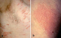
Fig. 1. (a) Digitiform parapsoriasis; (b) large plaque parapsoriasis.
Sampling
Once the clinical diagnosis was established, a histological examination was performed on each patient (the results were read by 2 independent pathologists). In addition, an immunochemical study (CD13) (by another physician) and a PCRγ/d denaturing gradient gel electrophoresis (DGGE) search for a T-cell lymphocytic clone in the skin and in the blood were performed. These examinations were performed on skin lesion biopsies obtained with a 4-mm punch. All skin biopsies were obtained from the same lesions.
Histology
Cutaneous biopsies were fixed in formalin, embedded in paraffin, dehydrated and then stained with haematoxylin-phloxine-saffron. Slides were classified into 2 groups, according to the aspect observed by the 2 pathologists:
1) Non-specific or eczematoid lesions or parapsoriasis image: the epidermis appeared normal, or spongiosis was seen, or an exocytosis of small lymphocytes groups in epidermis was present. A lympho-histiocytic perivascular infiltrate with no cellular atypia was observed in the superficial dermis. No atypical cells were present.
2) Images evoking epidermotropic CTCL: an epidermotropism was shown for mono-nucleated cells, with linearly arranged single lymphoid cells (closely apposed to basal keratinocytes and lined up along contiguous rete ridges like a necklace on the epidermal side of the dermal-epidermal junction). Cellular atypia, such as hyperchromatic and circonvoluted nucleus, were present. Cells were sometimes grouped in clusters with a typical aspect of “Pautrier’s abscesses”.
Immunohistochemistry
The immunological study was based on the analysis of the expression of the My7 antigen (CD13) in epidermal basal cells. Several studies (4, 5, 8) have shown that CD13 is expressed by 100% of cells in the normal or inflammatory skin, and that this expression disappears in the case of epidermotropic CTCL. Immunolabelling was performed on frozen cutaneous biopsies. Five-micron slides were prepared, then dried and fixed in acetone at 4°C.
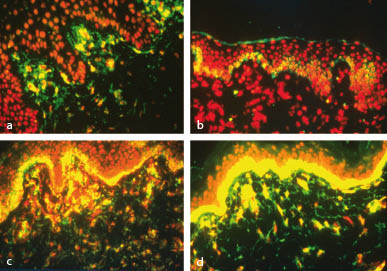
The My7 mouse monoclonal antibody (Immunotech, Marseille, France) was applied on the slides, after dilution to 1/20, for 30 min. As secondary antibody, an anti-mouse antibody was used (IgG Fab2) in combination with fluorescein (FITC), diluted to 1/15 in phosphate-buffered saline (PBS). A counter-colouration was finally performed with propidium iodide (150 µl iodide diluted in 4 ml of PBS). The results were read by a trained physician using a microscope. Results were given as the percentage of fluorescent basal cells (My7 +) in all basal epidermal cells (Fig. 2). Two subclasses were defined for reading: My7≤25% and My7 > 25%.
Fig. 2. Variable My7 (CD13) expression in basal epidermal cells (yellow fluorescence). (a) 0%; (b) < 25%; (c) ≥ 25%; (d) 100%.
Gene rearrangement
A study of the rearrangement of the TCR gene (T-cell receptor for the antigen) was performed using PCRγ/DGGE from frozen skin lesion biopsies for all patients, and, additionally, for 34/88 patients from blood samples. After extraction of the DNA from tissues, amplification was performed with a 480 Perkin Elmer Cetus thermocycler (40 cycles), and VγJγ primers were tested together (multiplex PCR). Amplification fragments were then separated by DGGE with a 10–60% gradient. Migration was performed at 170 V for 6 h. The gel was then stained for 30 min in an ethidium bromide bath and photographed under UV light.
Statistical methods
The statistical study consisted in the determination of a correlation between the studied factors. In this regard, the Mann-Whitney and χ2 tests were used, as well as the Fisher's exact test.
RESULTS
Demographic and clinical results
The study population comprised 88 patients (62 men, 26 women) with a mean age of 53.1 years at the onset of symptoms (age range 15–88 years), which most frequently occurred during the sixth or seventh decade. Of these 88 patients, 20 presented with a clinical aspect of digitiform PP and 68 with a large plaque PP. The sex ratio was much higher in the digitiform group (17 men, 3 women) compared with the other group (45 men, 23 women). Four patients (2 men, 2 women) in the large plaque group presented with a poikilodermal PP. Two of them were younger than 30 years at the time of diagnosis.
Histological results
A histological aspect of epidermotropic CTCL was found in 19/88 (21.6%) cases of PP, 16 in large plaque PP (23.5%) and 3 in digitiform PP (15%). Two aspects of MF were found in the poikilodermic PP (50%).
Immunological results
Expression of the My7 antigen was negative or < 25% for 53/88 (60.2%) of biopsies (12 in digitiform PP and 41 in large plaque PP). Disappearance of My7 was associated with the histological image of epidermotropic CTCL in 15/19 (78.9%) histological aspects of CTCL (p = 0.069) (Table I).
Table I. Histological features vs. My7 expression
| My7 Histology | 0–<25% | 25–100% | Total | |
| Non-specific PP | 38 (55%) | 31(45%) | 69 (100%) | |
| Epidermotropic CTCL | 15 (79%) p = 0.069 | 4 (21%) | 19 (100%) | |
| Total | 53 | 35 | 88 |
PP: parapsoriasis; CTCL: cutaneous T-cell lymphoma
Rearrangement of the TCR gene
Skin. No gene rearrangement was shown in any of the 20 patients who presented with digitiform PP. Among the 68 patients with large PP, 7 (10.3%) had a monoclonal gene rearrangement. For 4 patients it was associated with histology of early MF and for 3 with non-specific histology combined with disappearance of My7 antigen expression. A significant correlation was shown between the presence of a T-cell clone and a histological image of epidermotropic CTCL (p = 0.03) (Table II). No correlation was found between the presence of a T-cell clone in the skin and the disappearance of My7 antigen expression (Table III).
Table II. Gene rearrangement in the skin vs. histology
| Gene rearrangement Histology | Polyclonal | Monoclonal | Total | |
| Non-specific PP | 66 | 3 | 69 (100%) | |
| Epidermotropic CTCL | 15 (79%) | 4 (21%) p = 0.03 | 19 (100%) | |
| Total | 81 | 7 | 88 | |
| PP: parapsoriasis; CTCL: cutaneous T-cell lymphoma | ||||
Blood. The assessment was performed for 34 patients out of 88. Two circulating clones were found, for 2 patients with large plaque PP. In both cases, the histology showed an epidermotropic CTCL, but the gene rearrangement was negative in the skin.
Table III. Gene rearrangement in the skin vs. My7 expression
| Gene rearrangement My7 | Polyclonal | Monoclonal | Total | |
| 0–25% | 49 | 4 p = 1 | 53 | |
| 25–100% | 32 | 3 | 35 | |
| Total | 81 | 7 | 88 |
DISCUSSION
The recent validated criteria for early MF are persistent and/or progressive patch/thin plaques, non-sun-exposed location, size/shape variations and poikiloderma (3). At the clinical level, however, the differential diagnosis between grande plaque PP and early MF often remains difficult even for the dermatologist. At the histological level, several biopsies performed in a same plaque of PP may show different aspects with, in some places, an inflammatory non-specific lesion and, in other places, an aspect of early MF. Thus, the histology is not always significant enough for differentiating PP and early MF, knowing that about 20% of large plaque PP will evolve into a CTCL.
In our study we found a histological aspect of early MF in 19/88 PP (21.6%) and in 2/4 (50%) poikilodermic forms of PP. In addition, 3 digitiform PP (15%) had a histological picture of early MF, which is rarely reported (9–11). This is, however, in accordance with the study of King-Ismaël & Ackermann, who observed images of early MF while re-assessing their slides of digitiform PP (12).
Thus, it confirms that, at the clinical level, the differential diagnosis between PP and early MF remains difficult and that histology is not a sufficient criterion (13–16).
Concerning the interest in My7 antigen (CD13) to help in the diagnosis of early MF, in our series we observed that the expression of this antigen disappeared, mainly when the histological aspect was that of an epidermotropic CTCL, which is in agreement with former studies that showed a disappearance of the expression of the My7 antigen in cutaneous lymphomas (MF and Sézary’s syndrome) (4, 5). However, in our study the expression of this antigen also disappeared in 38 patients with PP with a non-histological aspect of MF. Thus, we did not find a correlation between the absence of My7 expression and a histological aspect of early MF. However, one hypothesis could be that the disappearance of My7 antigen occurs before the histological aspect of MF appears, raising the possibility of My7 as a prognostic marker of evolution to epidermotropic CTCL, although this remains to be demonstrated. Consequently, it will be interesting in the future to follow-up patients with a non-specific histology and a negative My7, in order to confirm this hypothesis.
As regards the study of the rearrangement of the TCR gene in skin lesions, our results are similar to published data. It was polyclonal for digitiform PP and monoclonal for 10.2% of large plaque PP. In the literature the gene rearrangement is mainly polyclonal for digitiform PP (17–19), whereas it is monoclonal in 7.5–50% of large plaque PP, depending on the studies (6, 7, 18, 20, 21). Our data demonstrate the existence of a correlation between histology of early MF and the identification of a gene rearrangement in the cutaneous lesion. In the literature, a clone is detected in early MF, by PCR in 45% (6) to 71% (7) of cases.
Regarding haematological data, circulating clones were found for only 2 patients with grande plaque PP associated with histological images of epidermotropic CTCL, but with no correlation with the expression of My7 by epidermal basal cells or with the presence of a cutaneous clone. It is difficult to draw conclusions based on such a small sample. However, these results seem to disagree with a former study performed by Delfau-Larue et al.: the frequencies of monoclonal populations found in the blood of 119 patients with cutaneous lymphoma were similar for histological images of lymphoma, benign inflammatory or non-specific dermatoses (22, 23). Circulating clones were also found more frequently in patients aged more than 60 years (24). The 2 patients with circulating clones in our study were aged less than 60 years. However, the meaning of the presence of an isolated circulating clone is unknown.
Thus, considering these histological, immunological and molecular biology data, it appears that these 3 criteria may be of interest for the dermatologist to differentiate early MF and PP, specifically through T-cell rearrangement. Further studies will determine whether CD13 is an early maker of the evolution of PP to MF (25–28).
Acknowledgements
We thank Mrs Mireille Thollon and Mrs Madeleine Yviquel for their valuable technical assistance.
REFERENCES
1. Brocq L. Les érythrodermies pityriasiques en plaques disséminées. Rev Gen J Praticiens 1897; 11: 577–590.
2. Brocq L. Les parapsoriasis. Ann Dermatol Syphiligr 1902; 3: 433–468.
3. Pimpinelli N, Olsen EA, Santucci M, Vonderheid E, Haeffner AC, Stevens S, et al. Defining early mycosis fungoides. J Am Acad Dermatol 2005; 53: 1053–1063.
4. Dreno B, Bureau B, Stalder JF, Litoux P. My7 monoclonal antibody for diagnosis of cutaneous T cell lymphoma. Arch Dermatol 1990; 126: 1454–1456.
5. Celerier P, Bureau B, Litoux P, Dreno B. Keratinocyte-lymphocyte interaction in cutaneous T-cell lymphoma. Modulation of keratinocyte antigen My7 by a soluble factor produced by T lymphocytes. Arch Dermatol 1997; 133: 837–840.
6. Mielke V, Staib G, Boehncke WH, Duller B, Sterry W. Clonal disease in early cutaneous T-cell lymphoma. Dermatol Clin 1994; 12: 351–360.
7. Staib G, Sterry W. Use of polymerase chain reaction in the detection of clones in lymphoproliferative diseases of the skin. Recent Results Cancer Res 1995; 139: 239–247.
8. Jumbou O, N’Guyen JM, Tessier MH, Legoux B, Dreno B. Long-term follow-up in 51 patients with mycosis fungoides and Sezary syndrome treated by interferon Br J Dermatol 1999; 140: 427–431.
9. Bonvalet D, Colau-Gohm K, Belaich S, Civatte J, Degos R. Les différentes formes du parapsoriasis en plaques. A propos de 90 cas. Ann Dermatol Venereol 1977; 104: 18–25.
10. Lambert WC, Everett MA. The nosology of parapsoriasis. J Am Acad Dermatol 1981; 5: 373–395.
11. Lambert C. Premycotic eruptions. Dermatol Clin 1985; 3: 626–645.
12. King-Ismaël D, Ackerman AB. Guttata parapsoriasis/digitate parapsoriasis (small plaque parapsoriasis) is mycosis fungoides. Am J Dermatopathol 1992; 14: 518–530.
13. Civatte J, Belaich S, Bonvalet D, Morel P, Schnitzler L. Lesions non tumorales dermoepidermiques. Chapter 5. In: Civatte J 8editor). Histopathologie cutanée. Paris: Flammarion; 1982: p. 158–161.
14. Jones REJ. Questions to the editorial board and others authorities. Am J Dermatopathol 1986; 8: 534–545.
15. Shapiro PE, Pinto FJ. The histologic spectrum of mycosis fungoides/Sezary syndrome. Am J Surg Pathol 1994; 18: 645–667.
16. Smoller BR, Bishop K, Glusac E, Kim YH, Hendrickson M. Reassessment of histologic parameters in the diagnosis of mycosis fungoides. Am J Surg Pathol 1995; 19: 1423–1430.
17. Muche M, Lukowsky A, Heim J, Friedrich M, Audring H, Sterry W. Demonstration of frequent occurence of clonal T cells in the peripheral blood but not in the skin of patients with small plaque parapsoriasis. Blood 1999; 94: 1409–1417.
18. Klemke CD, Dippel E, Dembinski A, Ponitz N, Assaf C, Hummel M, et al. Clonal T cell receptor gamma–chain gene rearrangement by PCR-based Gene-Scan analysis in the skin and blood of patients with parapsoriasis and early stage mycosis fungoides. J Pathol 2002; 197: 34–354.
19. Haeffner AC, Smoller BR, Zepter K, Wood GS. Differentiation and clonality of lesionnal lymphocytes in small plaque parapsoriasis. Arch Dermatol 1995; 131: 321–324.
20. Kikuchi A, Naka W, Harada T, Sakuraoka K, Harada R, Nishikawa. Parapsoriasis en plaques: its potential for progression to malignant lymphoma. J Am Acad Dermatol 1993; 29: 419–422.
21. Simon M, Flaig M, Kind P, Sander C, Kaudewitz P. Large plaque parapsoriasis: clinical and genotypic correlations. J Cutan Pathol 2000; 27: 57–60.
22. Delfau-Larue MH, Petrella T, Lahet C, Lebozec C, Bagot M, Roudot-Thoraval, et al. Value of clonality studies of cutaneous T lymphocytes in the diagnosis and follow-up of patients with mycosis fungoides. J Pathol 1998; 184: 185–190.
23. Delfau-Larue MH, Laroche L, Wechsler J, Lepage E, Lahet C, Asso-Bonnet M, et al. Diagnostic value of dominant T-cell clone in peripheral blood in 363 patients presenting consecutively with a clinical suspicion of cutaneous T-cell lymphoma. Blood 2000; 96: 2987–2992.
24. Posnett DN, Sinha R, Kabak S, Russo C. Clonal populations of T cells in normal elderly humans: T cell equivalent to “benign monoclonal gammapathy”. J Exp Med 1994; 179: 609–618.
25. Fraser-Andrews EA, Woolford AJ, Russell-Jones R, Seed PT, Whittaker SJ. Detection of a peripheral blood T cell clone is an independent prognostic marker in mycosis fungoides. J Invest Dermatol 2000; 114: 117–121.
26. Burg G, Dummer R. Small plaque parapsoriasis is an “abortive cutaneous T cell lymphoma” and is not a mycosis fungoides. Arch Dermatol 1995; 131: 336–338.
27. Burg G, Dummer R, Nestle FO, Doebbeling U, Haeffner A. Cutaneous lymphomas consist of a spectrum of nosologically different entities including mycosis fungoides and small plaque parapsoriasis. Arch Dermatol 1996; 132: 567–572.
28. Ackerman AB. If small plaque parapsoriasis is a cutaneous T cell lymphoma, even an “abortive” one, it must be mycosis fungoides! Arch Dermatol 1996; 132: 562–566.

