Arianna Zangrilli, Marina Papoutsaki, Luca Bianchi, Miriam Teoli and Sergio Chimenti
Department of Dermatology, University of Rome “Tor Vergata”, Viale Oxford 81, IT-00133, Rome, Italy. E-mail: arianna.zangrilli@libero.it
Accepted January 21, 2008.


Arianna Zangrilli, Marina Papoutsaki, Luca Bianchi, Miriam Teoli and Sergio Chimenti
Department of Dermatology, University of Rome “Tor Vergata”, Viale Oxford 81, IT-00133, Rome, Italy. E-mail: arianna.zangrilli@libero.it
Accepted January 21, 2008.
Sir,
Dermatomyositis (DM) is a connective tissue disease that classically manifests several characteristic findings: Gottron’s papules, periorbital heliotrope rash, poikiloderma and reticular telangiectasies (1). However there exist a wide variety of atypical cutaneous features that have been observed on occasion. In a subset of affected patients, dermatomyositis is believed to be paraneoplastic syndrome (2).
We describe here a case of bullous dermatomyositis (DM) in a patient who developed a small-cell lung cancer with aggressive fatal course.
CASE REPORT
A 57-year-old Caucasian man was observed in our clinic with a widespread erythematous eruption, redness of the knuckles and periorbital oedema with prominent involvement of the upper eyelids. The cutaneous manifestations were associated with erosions of the oral mucosa and periungual telangiectasia. Furthermore, tense vesiculo-bullous lesions of different size were observed on the arms and one large bulla was present on the left knee (Fig. 1). A concomitant swelling and tenderness of the joints associated with weakness, and strength reduction of proximal muscles were noted following clinical examination. Laboratory findings were remarkable for a white blood count of 15,500 cell/mm3 (normal 3600–11,500 cell/mm3), LDH of 627 U/l (< 430 U/l), CK 892 UI/l (10–190 UI/l), ANA titre of 1:320 with a speckled pattern, erythrocyte sedimentation rate (ESR) of 25 mm/h, CPK-MB 43 UI/l (0–24), myoglobin of 394.7 ng/ml (23–72 ng/ml), troponin T of 0.15 ng/ml (0.1 ng/ml), carcinoembryonic antigen (CEA) of 16.78 ng/ml (< 10 ng/ml). The diagnosis of bullous DM, a rare clinical variant of DM, was made through the histological examination and in accordance with the electromyography (EMG) results. In order to exclude the possible association of DM with malignancy, the following investigations were undertaken: chest radiography, upper and lower gastrointernal endoscopy, abdominal echography and total body computerized tomography (CT), the results of which were negative. Nevertheless, taking into consideration the possible association of DM, in particular the bullous variant, with malignancy, immunosuppressive protocols were avoided and high-dose intravenous immunoglobulins therapy (400 mg/kg for 5 days monthly) was instituted. Even though a rapid improvement of the cutaneous lesions, muscle weakness and laboratory findings were observed already after the first cycle of therapy, the patient showed a progressive and significant weight loss (10 kg in 2 months). Chest radiography and total body CT were then repeated and pulmonary lesions compatible with a lung carcinoma were detected. Shortly after this diagnosis, the patient died and the autopsy revealed a pulmonary invasive, poorly differentiated, small-cell lung carcinoma with bone metastases.
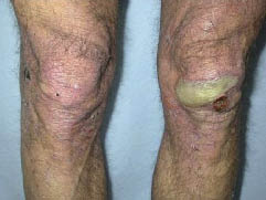
Fig. 1. One bulla eruption localized on the left knee.
DISCUSSION
DM is a rare progressive autoimmune disease characterized by inflammatory myopathy and typical skin lesions (1). Cutaneous manifestations commonly include Gottron’s papules, reticular erythema and telangiectasia, periorbital oedema with a heliotrope rash, papulosquamous eruption of the hairline, face and trunk, as well as poikiloderma. Vesicles and bullae have been rarely observed (2). The association of DM and malignancy has been well-documented since the original reports in 1916 (3). Therefore, in a subset of patients, DM is believed to be a paraneoplastic syndrome (4). The incidence rate of DM associated with cancer in adults varies between 6% and 60% (5) and in some cases the bullous DM variant seems to be predictive of the paraneoplastic nature (6). In a recently published pooled analysis of DM cases, a significantly increased incidence of several different malignancies, namely ovarian, lung, pancreatic, stomach, colon-rectal cancer as well as non-Hodgkin’s lymphoma, was reported (5). Although few references concerning vesicular and bullous lesions in DM can be found, this clinical variant is more likely related to internal malignancies (6). To the best of our knowledge, this is the first description of vesiculo-bullous DM associated with a small-cell lung carcinoma.
In our patient, the diagnosis of DM was based on the cutaneous lesions, although atypical, the muscle involvement, the laboratory tests, the cutaneous biopsy and the EMG. Nevertheless the association with malignancy was determined only some months after the diagnosis of DM. When the first diagnostic examination for internal malignancy is negative and treatment does not sufficiently eliminate symptoms, a new general examination is mandatory after 6–12 months. This case emphasizes the importance of early diagnosis of vesiculo-bullous DM in order to institute appropriate treatment and to perform accurate systemic evaluation for associated malignancies.
References
1 McCollough ML, Cockerell CJ. Vesiculo-bullous dermatomyositis. Am J Dermatophatol 1998; 20: 170–174.
2 Kaufmann R, Greiner D, Schmidt P, Walter M, Wolter M. Dermatomyositis presenting as plaque-like mucinosis. Br J Dermatol 1998; 138: 889–882.
3 Sterz G. Verhandlungen Arzlicher Gesellschaften: Berliner Klinische Wochenschrift 1916; 53: 488–489.
4 Wakata N, Kurihara T, Saito E, Kinoshita M. Polymyositis and dermatomyositis with malignancy: a 30-year retrospective study. Int J Dermatol 2002; 41: 729–734.
5 Hill CL, Zhang Y, Sigurgeirsson B, Pukkala E, Mellemkjaer L, Airio A, et al. Frequency of specific cancer types in dermatomyositis and polymyisitis: a population-based study. Lancet 2001; 357: 96–100.
6 Gentina T, Arbion F, Bernard A, Strecker A. Dermatomyositis and small-cell lung cancer: fortuitus association or paraneoplastic syndrome? Rev Pneumol Clin 2000; 56: 209–212.

