Nahide Onsun1, Ozlem Su1*, Yasemin Balsever Kural1, Yeliz Erdemoglu1, Nesimi Buyukbabani2, Cuyan Demirkesen3 and Yildiz Aydin4
1Department of Dermatology, Medical Faculty of Bezmialem Vakif University, Adnan Menderes Bulvarı Fatih, 2Department of Pathology, Istanbul Faculty of Medicine, 3Department of Pathology, Cerrahpasa Faculty of Medicine, and 4Department of Haematology, Cerrahpasa Faculty of Medicine, University of Istanbul, Istanbul, Turkey. E-mail: nonarir2011@hotmail.com
Accepted December 8, 2010.
Primary cutaneous B-cell lymphomas (CBCLs) are divided into three main types according to the WHO-EORTC classification: marginal zone B-cell lymphoma (MZL), follicle centre lymphoma (FCL), and diffuse large B-cell lymphoma, leg-type (DLBL) (1–4). A new tumour, lymph node, metastasis (TNM) staging system (Table SI (available from: http://www.medicaljournals.se/acta/content/?doi=10.2340/00015555-1108)) for CLs other than mycosis fungoides and Sezary syndrome was recently proposed. The new system provides documentation of disease extent/distribution and can be used to select appropriate treatment (3, 5).
The aims of our study on 10 CBCL cases were: (i) to characterize the clinicopathological features of the three subtypes of CBCL; (ii) to assess the value of the new TNM staging system; (iii) to evaluate response to the various treatment modalities and (iv) to assess the prognosis of CBCL with tumour staging.
PATIENTS AND METHODS
We reviewed the cases of 10 patients who received a diagnosis of CBCL between 1997 and 2007. These were classified according to the WHO-EORTC classification for CLs (Table SI). All patients had undergone complete staging, including complete blood cell count, chemistry panel, ultrasound examination of lymph nodes, and computed tomography scans of chest, abdomen, and pelvis, as well as bone marrow biopsy, as suggested by the ISCL-EORTC (5). Histological review was performed on haematoxylin-eosin stained sections, immunohistochemical staining including B-cell markers (CD20, CD79a), and at least one T-cell marker (usually CD3, CD5 or both), with additional staining for bcl2, bcl6, CD10, MUM-1 (in DLBCL). Positivity for bcl2, bcl6 on 1 MUM-1 was defined as expression of those markers by at least 50% of the tumour cells.
Medical records were reviewed for demographic data, localization, number, and extent of lesions at the time of diagnosis, according to the new TNM staging scheme (5) and for diagnosis according to the WHO-EORTC classification for CLs (1, 4). We reviewed serological tests for Borrelia infection, treatment modalities and response.
RESULTS
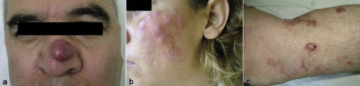
The CBCL patients were classified as MZL (n = 4), FCL (n = 3) and DLBL (n = 3) (Table I). The MZL patients TNM stages were T1a, T2a and T2b (mean age 49 years). The predilection sites for MZL were the head-neck (n = 2) and upper back (n = 2). Clinically, MZL began with pink, red, or red-brown nodules (Fig. 1a). The patients were treated with surgical excision or intralesional or systemic interferon (IFN)-α.Complete remission (CR) was achieved in all patients with MZL. Extracutaneous spread was observed in only one patient (T1a). The patient who refused surgical excision developed nodal metastases, showing transformation to large cell lymphoma and bone marrow involvement 21 months after the treatment. She then received eight cycles of combined chemotherapy with CHOP (cyclophosphamide, doxorubicin, vincristine, and prednisone). However, 8 months after the treatment, a cervical lymphadenopathy consistent with MZL was seen. She refused any further CHOP and was followed up without systemic involvement for 4 months (6). No recurrence was observed during the follow-up period of 24 months in three of the four patients (Table I).
Table I. TNM stage, treatment modalities, outcome and relapses of 10 investigated patients with cutaneous B-cell lymphoma
| Age/sex | CTCL subtype | T stage | Treatment | Outcome | Relapse | Re-treatment | Follow-up (months) |
| 50/M | MZL | T1a | Surgical excision | CR | – | – | 36 |
| 60/F | MZL | T1a | IL IFN-α (2a) | CR | + | CHOP | 72 |
| 36/M | MZL | T2a | IL IFN-α (2a) | CR | – | – | 12 |
| 50/M | MZL | T2b | S IFN-α (2b) | CR | – | – | 24 |
| 48/M | FCL | T2a | Surgical excision | CR | – | – | 24 |
| 34/F | FCL | T2b | RT, S IFN-α 2(b), C, M, I CHOP, R | NR | – | – | 6 |
| 17/M | FCL | T3b | S IFN-α (2b) | CR | + | R | 60 |
| 66/M | DLBL | T2b | CHOP | CR | – | – | 24 |
| 78/F | DLBL | T3a | CHOP | LFU | ? | ? | 1 |
| 67/M | DLBL | T3b | R | CR | – | – | 24 |
MZL: marginal zone B-cell lymphoma; CHOP: chemotherapy with cyclophosphamide, doxorubicin, vincristine, and prednisone; FCL: follicle centre lymphoma; DLBL: diffuse large B-cell lymphoma, leg-type; C: corticosteroids; CR: complete remission; I: imiquimod; IFN: interferon; IL: intralesional; LFU: lost to follow-up; M: methotrexate; NR: no response; R: rituximab; RT: radiotherapy; S, systemic.
Fig. 1. (a) Red nodule of marginal zone B-cell lymphoma on the nose. (b) Pink-red nodules and plaques on the left cheek of the patient with follicle centre lymphoma. (c) Violaceous nodules and plaques on the left leg of the patient with diffuse large B-cell lymphoma, leg-type.
The three FCL patients were T1a, T2b and T3b (Table I). The median age was 33 years. The predilection site for FCL was the chest (n = 2), but tumours were also found on head-neck (n = 1), upper back, and upper arms (n = 1). Clinically, FCL began with skin-coloured, pink, or red nodules and plaques (Fig. 1b). One patient with T1a had surgical excision, but another with T2b did not respond to any treatment modalities. The last patient with T3b was initially treated with systemic IFN-α-2b successfully, but local recurrence occurred after 36 months. The patient was re-treated with rituximab with excellent results.
The three DLBL patients were T2b, T3a and T3b (mean age 70.3 years). The predilection sites were the legs, but tumours were also found on the upper arms in one of the three patients. DLBL started with erythematous-to-violaceous nodules or plaques (Fig. 1c). Ulceration was observed in two of three patients. Two of the three patients (T2b, T3a) were treated with CHOP. The last patient with T3b was treated successfully with rituximab. CR was achieved in two of the three patients. Relapse or extracutaneous spread was not observed in these two patients during a mean follow-up time of 24 months. Serological tests for Borrelia infection, performed on all patients, were negative.
DISCUSSION
The prevalence of CBCL was only 7.5% of our total CLs. This is comparable with published literature (1, 2). The EORTC study found a female/male ratio of 2:1 (7), but some other studies showed male predominance, as in our study (3). To our knowledge, this is the fourth study of CBCL staged according to the new TNM staging system (3, 8, 9). Forty percent of our patients had MZL. This is slightly higher than in previously reported studies (2, 10), but consistent with the results of Golling et al. (3). The mean age was in the 50s, similar to that found in other reports (11).
IFN-α resulted in CR in two patients with MZL and in one patient with FCL, but recurrence was observed in the latter patient. Cozzio et al. (12), reported excellent results with IFN-α-2a with MZL, but relapses were observed in 25% of the patients. We suggest that intralesional IFN-α may be a valuable alternative to surgery and radiotherapy as a first-line treatment of MZL and FCL with T1 or T2 stage. We got no response to rituximab in one patient with FCL who was resistant to all treatments. Gellrich et al. (13) achieved 70% complete response with rituximab. Two patients with DLBL and one patient with MZL were treated with CHOP, but in the MZL patient nodal metastases were seen after the treatment (6). None of our 9 patients with CBCL had died of lymphoma after a median follow-up period of 31 months.
REFERENCES