


Jens Malte Baron1 and Bernhard Lüscher2
1Department of Dermatology and Allergology and
2Institute of Biochemistry and Molecular Biology
RWTH Aachen University,
Pauwelsstraße 30,
DE-52074 Aachen, Germany
In 2004, interleukin-31 (IL-31) was described as a short-chain 4-helix bundle cytokine that is expressed by activated CD4+ T cells, preferentially by T cells skewed toward a TH2 phenotype (1). Recently it has been demonstrated that human mast cells (MC) are also a source of IL-31 (2). Moreover, monocytes, macrophages, immature and especially mature monocyte-derived dendritic cells produce IL-31 in response to ultraviolet (UV) irradiation and hydrogen peroxide (H2O2) treatment (3). Low IL-31 mRNA expression levels have been detected in tissues from testis, bone marrow, skeletal muscle, kidney, colon, thymus, small intestine and trachea (1). In normal human epidermal keratinocytes and dermal fibroblasts-enhanced IL31 mRNA expression is measured upon H2O2 stimulation (3). In the skin of NC/Nga mice with scratching behaviour, an animal model of atopic dermatitis (AD), expression of IL-31 mRNA was significantly higher than that in NC/Nga mice without scratching behaviour. Together, these findings suggest that IL-31 is associated with TH2-driven pruritic skin diseases, and that IL-31 may participate in the cause for itch sensation (4, 5).
IL-31 signals through a heteromeric receptor complex composed of the IL-31 receptor alpha (IL-31RA) and the oncostatin M receptor beta (OSMR) subunits. Engagement of the receptor complex results in activation of Janus kinase (JAK) tyrosine kinases and subsequently of different signalling molecules, including different signal transducers and activators of transcription (STAT) factors, as well as the MAPK and PI3K signalling pathways (Fig. 1). These pathways are activated in various cell types, including lung epithelial or malignant melanoma cells (6–8).
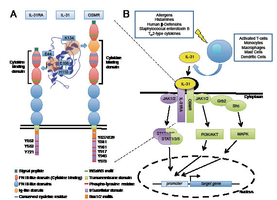
Fig. 1. Cytokine interleukin-31 (IL-31)-dependent signal transduction. (A) The IL-31 receptor is composed of IL-31 receptor alpha (IL-31RA) and oncostatin M receptor beta (OSMR). The domain structure of the two receptor subunits is indicated. Both have a C-terminal signalling peptide that is cleaved off during biosynthesis and translocation of the protein into the endoplasmic reticulum. IL-31 binds first to the IL-31RA (through two fibronectin III-like domains) and subsequently to the OSMR (through two fibronectin III-like and an immunoglobulin-like domain). Amino acids demonstrated to be important for binding are indicated; for binding to IL-31RA E44, E106, and H110; for binding to OSMR K134. The tyrosine residues that are phosphorylated by JAK kinases and mediate signalling are indicated. (B) Different cell types produce IL-31, as indicated; in many situations IL-31 production is regulated by different stimuli, including allergens, superantigens, and cytokines. The signalling that is stimulated is the JAK-STAT, the MAPK (i.e. ERK, p38 and JNK kinases), and the PI3K-AKT pathways. Of note is that IL-31RA activates predominantly different signal transducers and activators of transcription (STAT) molecules (purple), whereas OSMR is capable of stimulating all three pathways (green). These control downstream effectors, including transcriptional regulators that mediate and control IL-31-dependent target gene expression.
Expression of IL-31Rα and OSMRβ mRNA can be induced in activated monocytes, while their expression is constitutive in skin, testis and thymus, arguing that these organs are probably responsive to IL-31 (1, 9, 10). IL-31RA and OSMRβ are also found co-expressed in a subset of neurons of murine and human dorsal root ganglia (9, 11), which represents the site where the soma of cutaneous sensory neurons are located, while their sensory fibres protrude directly into the skin. These sensory neurons might be implicated in the sensing of itch and are possibly stimulated by IL-31. Recent studies demonstrated variations of IL-31RA expression in normal human epidermal keratinocytes that are dependent on the status of cellular differentiation and influenced by pro-inflammatory cytokines such as interferon-gamma (8).
While enhanced IL-31 mRNA expression has previously been detected in skin samples of inflammatory skin diseases such as AD, allergic contact dermatitis or prurigo nodularis (9, 12, 13), hardly anything is known about the protein expression of this cytokine in lesional skin from patients with inflammatory skin diseases. This question has now been addressed in an article in this issue of Acta Dermato-Venereologica by Nobbe and colleagues (p. 24–28) (14).
In their study they performed immunohistochemical staining for IL-31, IL-31RA and OSMR in formalin-fixed paraffin-embedded biopsy specimens of TH1- and TH2-weighted, pruritic and non-pruritic skin diseases. The authors demonstrate an enhanced IL-31 expression in biopsies of fresh lesions from patients with the extrinsic type of AD compared with controls. In 87% of samples from patients with AD cytoplasmic immunoreactivity for IL-31 was present in cells infiltrating the dermis, in contrast to epidermal cells, in which no significant staining could be detected (14). Comparisons of immunoreactivity in the dermal infiltrate of the examined itching skin diseases (AD, pruritus sine materia, psoriasis inverse, prurigo nodularis, Sézary syndrome, notalgia paraesthetica) revealed increased IL-31 immunoreactivity only in AD. Similar results were obtained by comparing different TH2-weighted diseases (AD, localized alopecia areata, mycosis fungoides, Sézary syndrome), showing increased IL-31 immunoreactivity only in samples from subjects with AD.
These studies confirm, at the protein level, the relationship between IL-31 expression and AD that was previously identified at the mRNA level. However, the presented data do not support a general relationship between IL-31 protein expression and pruritic or TH2-mediated diseases (14). Therefore IL-31 might play a role in the pathogenesis of AD and presents a putative therapeutic target, especially in the acute phase of this disease.
ACKNOWLEDGEMENTS
We would like to thank Christian Cornelissen for assisting in preparing Fig. 1. Work in the authors’ laboratories has been supported by a grant of the Deutsche Forschungsgemeinschaft (SFB 542 project C11).
REFERENCES

