


Pain and Hyperpigmentation of the Toes: A Quiz
Felipe Maurício Soeiro Sampaio1, Vitor Ribeiro Gomes de Almeida Valviesse1, Julia Ocampo Lyra-da-Silva2, Emerson Cicilini Mesquita1, Luciana Gomes Pedro Brandão1 and Antonio Carlos Francesconi do Valle 1
1Oswaldo Cruz Foundation, Marino da Costa Street, 217. Compl. 201. Ilha do Goverador, 21940-210 Rio de Janeiro, and 2Bonsucesso Federal Hospital, Rio de Janeirio, Brazil. E-mail: felipemauricio@uol.com.br
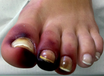
A 20-year-old female student, living in Rio de Janeiro, Brazil, developed well-delimited, asymptomatic, dark-reddish macules, on the distal phalanxes of the 3 first toes of her left foot (Fig. 1). The lesions appeared minutes after having put her shoes on, with an immediate sensation of burning. The shoes had been left outdoors overnight and she had observed an animal inside.
The patient appeared to be in a good general state of health, with no palpable lymph nodes or vascular involvement. There were no signs and symptoms of systemic disease. Diagnosis was based on epidemiological factors, clinical examination and observation of the agent.
Fig. 1. Clinical presentation of the skin lesions: well-delimited, asymptomatic, dark-reddish macules, on the distal phalanxes of the 3 first toes of the left foot.
What is your diagnosis? See next page for answer.
doi: 10.2340/00015555-1645
Pain and Hyperpigmentation of the Toes: Comment
Acta Derm Venereol
Diagnosis: Hyperpigmentation of the toes caused by millipedes
Millipedes are arthropods, of the Diplopoda Class (1.3). They are long in form, segmented, with 2 pairs of feet on each segment, except for the first 4 segments and the head. Millipedes are found in dark, humid places and consume decaying vegetal material (1). In Brazil, the most important species is the Rhinocricus padbergi (1). Due to their phenotypic characteristics, they are often mistaken for earwigs or centipedes.
Liquids and vapours expelled by the arthropod as a defence mechanism can cause burning of the skin and skin hyperpigmentation in humans. Discharge of these fluids, promotes an unpleasant smell reminiscent of urine. The fluids are irritating and contain benzoquinone compounds, which are responsible for the hyperpigmentation (1–6). However, reported cases of people injured by millipedes are rare. Arab et al. (7) found the 2 most volatile constituents: 2-methyl-1,4-benzoquinone and 3.3 to 4.5-tetrahydro-1H-pyrrolo-[2.3-b]pyridine-2.6-dione in the arthropod’s glands, to be responsible for the hyperpigmentation. Prolonged contact could create vesicles, blisters and ulceration (5). Ocular accidents cause conjunctival erythema, tearing, burning and, rarely, corneal ulceration (6).
Diagnosis is reached through epidemiological history and clinical examination. As there is no specific laboratory examination, finding the arthropod is essential for diagnosis.
Knowledge of this entity among clinicians is fundamental in differential diagnosis, as there are clinical similarities with systemic diseases, such as meningococcaemia, bacterial endocarditis, cryoglobulinaemia and collagenosis. Arterial ischaemia (2), traumas and child abuse must also be eliminated as differential diagnostic.
In spite of this worrisome clinical presentation, intense washing of the affected area of skin with soap and water or alcohol is the only recommendation (1–3). Hyperpigmentation disappears in weeks or months, requiring no topical depigmentation treatment. Topical corticosteroids could be used in symptomatic cases that display burning and vesiculation (1).
REFERENCES

