


Andrea Diociaiuti1, Alessandro Inserra2, Irene Fuertes De Vega3, Cristina Rota1, Tiziana Surrenti1, Loredana Giraldi1, Maria Rosaria Piemontese4, Isabella Giovannoni5, Francesco Callea5 and May El Hachem1
1Dermatology Unit, 2General and Thoracic Surgery Unit, 5Department of Pathology and Laboratory Medicine, Bambino Gesù Children’s Hospital, IRCCS, IT-00165 Rome, Italy, 3Dermatology Unit, Hospital Clínic, Barcelona, Spain, and 4Medical Genetics Unit, “Casa Sollievo della Sofferenza” Hospital, IRCCS, San Giovanni Rotondo, Italy. E-mail: andrea.diociaiuti@tin.it
Accepted Apr 30, 2014; Epub ahead of print May 9, 2014
Naevoid basal cell carcinoma syndrome (NBCCS), also known as Gorlin syndrome, is a rare, autosomal dominant disorder with systemic involvement (1). It is caused by germline mutations in the PTCH1 gene (locus 9q22.3–q31), which produce a tumour suppressor protein (2).
The prevalence of NBCCS varies from 1/30,000 to 1/256,000 (3), depending on the geographic location.
NBCCS is characterised by multiple basal cell carcinomas (BCCs) which usually develop at a young age, palmar or plantar pits, odontogenic keratocysts that appear in the first, second and third decades (4) of life and ectopic calcification of the falx cerebri. However, many other anomalies, such as macrocephaly, hypertelorism, cleft lip and/or palate, skeletal and eye abnormalities have been described in patients with NBCCS. Several low-frequency neoplasms, such as medulloblastoma, meningioma, ovarian and cardiac fibroma have also been reported in these patients.
The diagnosis of NBCCS is based on clinical findings and familial history of the patient and can be made if 2 major and 2 minor criteria are met (4). Genetic counselling must be considered (3). Furthermore, during pregnancy, ultrasound scans can be performed to detect developmental malformations.
Patients with NBCCS usually present to a dermatologist because of skin lesions; however, in addition to a periodic dermatological evaluation, management of the disease may require a wide range of specialists such as dentists, cardiologists, oncologists, and orthopaedic surgeons.
Case report
A 22-month-old girl with a 2-month history of a thoracic cage mass (Fig. 1A) was referred to our institution for investigation and treatment of this lesion. Prior to surgery, she was referred to the dermatology unit for the evaluation of several cutaneous lesions previously diagnosed as melanocytic naevi.
At physical examination she showed multiple small, pigmented papules involving the face, trunk (Fig. 1D), and limbs. Macrocephaly, moderate hypertelorism (Fig. 1B) and palmar pits (Fig. 1C) were also observed. Furthermore, the patient’s mother presented with multiple facial lesions clinically consistent with BCCs, and reported that in the past she had undergone several excisions of BCCs and that she had been diagnosed as suffering from NBCCS.
Whole body CT scan showed no alterations in the CNS and ribs but revealed the presence of a solid oval shaped mass measuring 7 × 4 × 2 cm in the thoracic right cage with evident enhancement after contrast. The thoracic mass was excised. Histological examination showed proliferation of rhabdomyoblasts resembling either ganglion cells with vesicular nuclei and prominent nucleoli, or ribbon or strap-like cells with deeply eosinophilic cytoplasm and cross striations. Both types of cells had a benign appearance. These findings were consistent with fetal rhabdomyoma (Fig. S1D1).
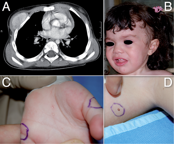
Fig. 1. Solid oval-shaped hyperintense mass in the thoracic cage visible on CT scan (A). Facial view of the patient showing increased head circumference, broadened nasal root, frontal bossing and mild hypertelorism (B). Numerous palmar pits on the left hand (C). Basal cell carcinoma on the trunk with “naevoid” appearance (D). A written permission is given to publish this figure.
A total of 7 cutaneous lesions were also removed (one on the face, 4 on the trunk and 2 on the left hand). These lesions were detected by dermoscopy that revealed blue-gray globules inside all the lesions, as described by Feito-Rodríguez et al. (5), except for one on the left hand that had a round shaped cystic appearance. Histology analysis revealed 5 lesions that appeared to be basal cell carcinomas (Fig. S1A and B1) corresponding to a tumour nodule located in the deep dermis. The nodule was encapsulated and composed of microfollicles lined by cuboidal or basaloid cells. Despite the absence of obvious peripheral palisade arrangement, the tumour was classified as a basaloid tumour with unusual appearance that can occur in NBCCS (Fig. S1C1).
During the subsequent follow-up the disease was particularly aggressive and the patient underwent 2 further surgical interventions with CO2 laser vaporisation, or surgical removal of hundreds of small BCCs.
The patient was diagnosed as having an NBCCS based on the presence of 3 major (palmar pits, first degree relative with Gorlin syndrome, multiple BCCs) and one minor criteria (macrocephaly).
To confirm the diagnosis of Gorlin syndrome a molecular study was performed on the child and the mother. All 23 coding exons of the PTCH1 gene (GeneBank accession number: NC_000009.10) were amplified by PCR and sequenced. A nucleotide change c.585-1G>A in the splice acceptor site of intron 3 (Fig. S2A1) was identified in both the child and the mother. RT-PCR analysis, carried out on an RNA sample from the patient using the High-Capacity cDNA Reverse Transcription Kit (Applied Biosystems, Foster City, CA) allowed us to assess the effect of the c.585-1G>A mutation at the RNA level and revealed the skipping of exon 4 (Fig. S2B1).
Discussion
To the best of our knowledge, there have been no reports in the literature concerning NBCCS patients younger than ours. In patients with NBCCS the mean age of BCCs onset is 20 years as reported by Shanley et al. (6). Even if BCCs have been previously described in the paediatric population not only in association with NBCCs, but also with Bazex syndrome, solid organ transplants, albinism and xeroderma pigmentosum (7, 8).
Patients with NBCCS must avoid unprotected sun exposure (4). Diagnostic and management protocols were recently proposed in order to limit morbidity and mortality in the affected patients (9)
Surgical excision, electrodessication and curettage are performed to treat small BCCs. Other treatments such as laser ablation, photodynamic therapy, and topical chemotherapy may be effective in patients with multiple lesions (10). Imiquimod 5% cream also seems to be useful (11). Total body application of topical 0.1% tretinoin cream, has been successfully used in patients with superficial BCCs (12).
LDE225 is a recently developed selective smoothened human and murine antagonist that is normally controlled by the PTCH1 gene. Treatment of NBCCS patients with 0.75% LDE225 cream results in a complete or partial regression of BCC without any side effects, offering promising therapeutic options (13).
In our patient additional tumours were a thoracic foetal rhabdomyoma and an unusual skin tumour, with a basaloid component, mimicking an eccrine adnexal tumour. The latter skin tumour was located in the deep dermis of the palm and was encapsulated and composed of microfollicles combined with cuboidal and basaloid cells.
Foetal rhabdomyoma is an extremely uncommon benign neoplasm with skeletal muscle differentiation that has already been reported in association with NBCCS (14). Quite recently a thoracic foetal rhabdomyoma has been reported in association with the mutation c.585-1G>A (15), as in our case.
The authors declare no conflict of interest.

1http://www.medicaljournals.se/acta/content/?doi=10.2340/00015555-1892
References


