


Giovanni Ghigliotti1, Giovanni Di Zenzo2, Emanuele Cozzani1*, Franco Rongioletti1, Elena De Col1, Carlotta Pastorino1, Giovanni Murialdo3 and Aurora Parodi1
1Sezione di Dermatologia, 3Clinica di Medicina Interna 2, IRCCS AOU San Martino- IST, Di.S.Sal, University of Genoa, 2Laboratorio di Biologia Molecolare e Cellulare, IDI - IRCCS, Rome, Italy. *E-mail: emanuele.cozzani@unige.it
Accepted Jun 11, 2015; Epub ahead of print Jun 15, 2015
Paraneoplastic pemphigus (PNP), described in 1990 by Anhalt et al. (1), is an autoimmune mucocutaneous blistering disease characterized by severe mucosal erosions and polymorphic cutaneous lesions. In 2001 the term paraneoplastic autoimmune multi-organ syndrome (PAMS) was coined (2) to reflect its frequent multi-organ involvement and numerous clinical variants (pemphigus-like, pemphigoid-like, erythema multiforme-like, graft-versus-host disease-like, and lichen planus-like). This disease is most often associated with haematological malignancies, and has a high mortality rate (90%) with less than one-year mean survival.
We report here a case of PNP/PAMS that was associated with retroperitoneal Kaposi’s sarcoma (KS) and evolved through 3 different clinical variants (pemphigus-like, lichen planus-like and erythema multiforme-like). This is the first report of an association of PNP/PAMS with KS in literature.
CASE REPORT
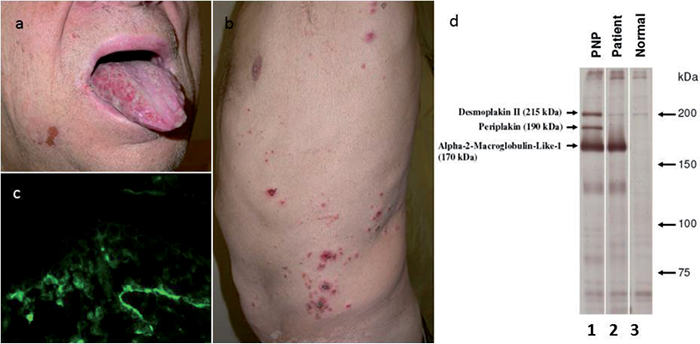
An HIV-negative 70-year-old man was treated with hydroxychloroquine (400 mg per day) for an erosive lichen planus involving the tongue, confirmed by histopathology and direct immunofluorescence (DIF). One month later, the patient showed a worsening of the oral ulcerations (Fig. 1a), and vesicles and blisters appeared on his trunk and lower limbs (Fig. 1b). Histopathology of a skin lesion revealed suprabasal intraepidermal blistering (not shown) and DIF showed intercellular deposition of IgG and C3 associated with C3 deposition at the basal membrane zone (BMZ) (Fig. 1c). The diagnosis of PNP was confirmed by indirect immunofluorescence (IIF) on rat bladder and by immunoprecipitation, with reactivity directed to the alpha-2-macroglobulin-like-1 (A2ML1) protein (Fig. 1d).
Fig. 1. (a) Ulcerations of the tongue. (b) Vesicles and blisters on the trunk. (c) Intercellular deposition and deposition of C3 at the basal intercellular substance associated with C3 deposition at the basal membrane zone. (d) Immunoprecipitation on radiolabelled cultured keratinocytes: Lane 1 (paraneoplastic pemphigus (PNP)): control PNP serum immunoprecipitating the 215 kDa desmoplakin II, the 190 kDa periplakin, and the 170 kDa alpha-2-macroglobulin-like-1; Lane 2 (Patient): serum of this patient immunoprecipitating the alpha-2-macroglobulin-like-1. Lane 3 (Normal): normal control serum showing no positive reaction.
Hydroxychloroquine was stopped and oral prednisone, at an initial dosage of 75 mg per day, was introduced with partial response. Extensive work-up, including an abdominal computed tomography (CT) scan, revealed a mesenteric mass in the peritoneum, below the diaphragm and next to the pancreas, with some enlarged lymph nodes, suggestive for a lymphoproliferative disorder. Despite treatment with glucocorticoids, the cutaneous blistering and oral erosions worsened. In addition, the patient developed many lilac itchy papules on the back and an erythematous purplish hyperkeratotic plaque on the inner surface of the left knee. Histopathology and DIF of a lilac papule on the patient’s back led to a diagnosis of lichenoid dermatitis with C3 and fibrinogen deposit in the cytoplasm of the keratinocytes and intercellular deposition of C3. Histopathology of the plaque on the knee was also consistent with lichenoid dermatitis. The patient’s general clinical condition worsened. He developed respiratory failure suggestive of bronchiolitis obliterans and erosive conjunctivitis associated with a polymorphous eruption of erythematous targetoid plaques localized on the upper extremities, together with a worsening of the oral erosions. Oral prednisone was increased to 125 mg/day. Fluoro-deoxyglucose positron emission tomography/computed tomography (PET-CT) (Fig. S1a, b1) showed intense tracer concentration in the peritoneal mass as well as in the thoracic and abdominal lymph nodes, spleen, right lobe of the liver, gastric area and, curiously, in the cutaneous plaque on the knee. Biopsy of the peritoneal mass revealed a malignant neoplasm composed of interconnected bands of medium-large sized spindle cells, with hyperchromatic, slightly pleomorphic nucleus, inconspicuous nucleoli and eosinophilic cytoplasm. In addition, dilated thin-walled vessels at the periphery of the tumour with lymphangioma-like areas were associated with an infiltrate of lymphocytes and plasma cells (Fig. S2a1). Immunohistochemical staining was positive for CD34, CD31 and human herpesvirus 8 (HHV-8) (Fig. S2b1), leading to a diagnosis of KS. The skin biopsy from the knee was re-evaluated and, below the lichenoid infiltrate, histological features of KS were detected. HHV-8 was also positive in some spindle cells adjacent to dilated vessel walls in the deep dermis, suggesting an early phase of KS. The retroperitoneal mass was considered inoperable, and chemotherapy with intravenous injection of vincristine 2 mg and cyclophosphamide 1 g once a week, with doxorubicin 60 mg 4 times a week, was introduced. After 3 days, the patient developed severe leukopaenia and septic shock (toxic shock syndrome) and eventually died. No post-mortem was performed.
DISCUSSION
A diagnosis of PNP/PAMS is based on 3 major criteria proposed by Schlesinger et al. (3); (i) polymorphic mucocutaneous eruptions, (ii) concurrent internal neoplasia, and (iii) serum antibodies reacting to plectin, desmoplakin I and II, bullous pemphigoid antigen I, envoplakin, periplakin or antigen of 170 kDa; and 3 minor criteria: (i) histological evidence of acantholysis, (ii) DIF showing intercellular and BMZ staining for IgG and complement, and (iii) IIF staining with rat bladder epithelium for circulating autoantibodies. Of these, PNP/PAMS patients should meet at least 3 major, or 2 major and 2 minor criteria. According to clinical PAMS criteria, our patient showed peculiar clinical features and developed 3 different mucocutaneous variants of the disease during its evolution: lichen planus-like lesions at the onset, then pemphigus-like lesions and, finally, erythema multiforme-like lesions with targetoid features (4, 5). Immunologically, our patient presented the classical criteria of PNP/PAMS with intercellular deposition of IgG and linear deposition of IgG and C3 at the BMZ. IIF on rat bladder transitional epithelium showed intercellular deposition of IgG autoantibodies, and immunoprecipitation revealed reactivity against the alpha-2-macroglobulin-like-1 protein, which is considered specific to PNP/PAMS. Furthermore, anti-A2ML1 autoantibodies appear to be associated with early onset of the disease (6).
PNP/PAMS is associated with different haemolymphoproliferative neoplasms (84%), such as non-Hodgkin’s lymphoma (38.6%), chronic lymphocytic leukaemia (18.4%) and Castelman’s disease (18.4%) (7). Solid neoplasms have rarely been reported in association with PNP/PAMS, with carcinomas in 8.6% of cases and sarcomas in 6.2% (8). Our patient is the first case of PNP/PAMS associated with KS to be described in the literature. Concerning the association with KS, our patient tested negative for HIV. The retroperitoneal sarcoma was diagnosed only a few weeks after starting prednisone and was thus unlikely attributed to drug immunosuppression. Moreover, an extensive work-up was negative for additional malignancies possibly inducing immunosuppression. The relationship between PNP/PAMS and the neoplasm is still unclear. In patients with thymoma, Castleman’s disease and dendritic cell sarcoma, a direct role of immune cells in the autoantibodies production has been hypothesized. In particular, PNP patients with Castleman’s disease, who had several tumour B-cell clones producing autoantibodies against desmosomal and hemidesmosomal proteins, showed a remarkable improvement in symptoms after removal of the tumour. Alternatively, the tumour proteins may cross-react with PNP epidermal antigens and induce the autoantibody production (7). Our case suggests that the tumour may also act as a stimulus for the immune system without being directly responsible for autoantibody production.
This report highlights the difficulty of diagnosing PNP/PAM in its early phase, with extreme polymorphism of the skin lesions, and demonstrate a noteworthy association with KS characterized by a lichenoid picture with early KS findings in the same cutaneous lesion.
ACKNOWLEDGMENTS
The authors would like to acknowledge the support of the Italian Ministry of Health (number RF2309790).

1http://www.medicaljournals.se/acta/content/?doi=10.2340/00015555-2169
REFERENCES


