


1”Onkoderma” – Policlinic for Dermatology and Dermatologic Surgery, 2Department “Medicinal Information and Non-interventional Studies”, Bulgarian Drug Agency, Sofia, Bulgaria, 3Department of Dermatology and Allergology, Academic Teaching Hospital Dresden-Friedrichstadt, Friedrichstrasse 41, DE-01067 Dresden, Germany, 4Department of Pathology, University of Virginia Health System, Charlottesville, USA, and 5Medical Institute of MVR – Ministry of Interior, Department of Dermatology, Venereology and Dermatologic Surgery, Sofia, Bulgaria. *E-mail: wollina-uw@khdf.de
Accepted Jun 1, 2016; Epub ahead of print Jun 15, 2016
Cutaneous epithelioid angiomatous nodule (CEAN) is a rare benign vascular proliferation within the spectrum of epithelioid/histiocytoid vascular lesions, characterized by histiocytoid endothelial cells, generally found in the dermis or subcutis and, rarely, in some visceral organs (1, 2). This group of vascular tumour-like lesions was identified by Rosai in 1979 and accommodates several cutaneous and extracutaneous diseases, each of which is characterized by the proliferation of vessels with peculiar “histiocytoid” endothelial cells (3). Brenn & Fletcher (4) set CEAN apart as a distinct entity in 2004. We are also of the opinion that CEAN should be considered a separate entity, although it is not yet listed in the International Society for Vascular Anomalies (ISSVA) classification (5).
Currently, CEAN is considered a reactive condition with rapid growth but a benign clinical course, and should be differentiated carefully from epithelioid haemangioendothelioma and epithelioid angiosarcoma (6). Although the aetiology of CEAN is not fully understood, no association with either infection, trauma, nor immunosuppression has yet been described (6). Associations with other conditions are rarely reported, but these include capillary malformation (CM) (7), breast cancer (8) and pyogenic granuloma (9).
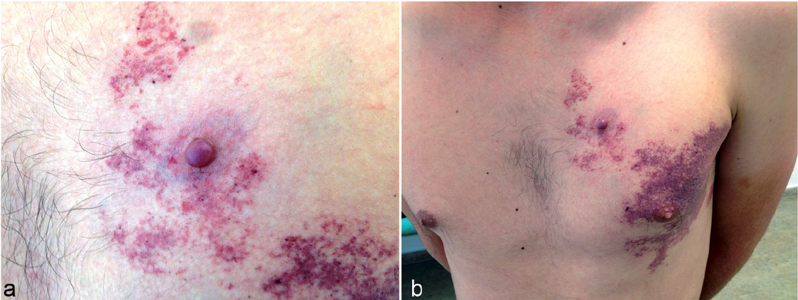
A 30-year-old Caucasian man consulted us with a tumorous formation arising in a pre-existing CM (port wine stain (PWS)-type). His family history was negative for dermatological diseases. A whole-body dermatological examination established a unilateral CM (PWS-type) along most of the skin of the left breast. The patient reported a reddish, oval-shaped, tumour with a smooth surface, which had enlarged rapidly during the last year, measuring approximately 12 mm in diameter, located on the skin in the region of the left breast (Fig. 1). A diagnosis of CEAN arising in CM (PWS-type) was confirmed histopathologically after surgical excision (Fig. 2).
Fig. 1. Clinical mani-festation of cutaneous epithelioid angiomatous nodule (CEAN), associa-ted with unilateral capillary malformation (CM) (port wine stain (PWS)-type). (a) CEAN (histiocytoid haemangioma) in CM (detail). (b) Overview
Fig. 2. Histopathological examination of cutaneous epithelioid angiomatous nodule (CEAN). Venular and cavernous types of blood vessels, with solid cell spaces, represented by spindle-like and epithelioid-like cells. The spindle-like cells form a network of fissures spaces and short sheaves (hematoxylin-eosin stain; original magnification × 36).
Abnormal dilated vessels were identified around the CEAN in the dermis.
Neurological examination and mental health assessment did not reveal any pathology; neither did pulmonary examination, abdominal ultrasound or Doppler echocardiography. No enlarged lymph nodes were observed. EMG examination revealed normal conduction of the peripheral nerves of the pectoral muscles and no difference in the latency time was discovered. Blood tests were within the normal range. Laser therapy was planned as a therapeutic option for the nevus flammeus in the future. Other vascular lesions of importance in differential diagnosis were excluded.
The simultaneous coexistence of CEAN and CM is an extraordinary finding. CEAN occurs most commonly as a rapidly-growing, reddish, nodule or papule, in the head-and-neck region of adult patients, equally distributed among males and females (6). Endothelial cells express CD31 and CD34, while pericytes are positive for smooth muscle actin (SMA) (7). Although usually presenting as a solitary lesion, multifocal presentation has been also reported, with an unusual predilection for the glans penis and forearm (6, 10). The first description of CEAN arising in pre-existing vascular malformation (CM) was reported in 2011 (7). The authors proposed CEAN as a variant of pyogenic granuloma with a mostly epithelioid appearance, developed in pre-existing CM (7). PWS is a congenital cutaneous capillary-venous malformation due to mutations of guanine nucleotide-binding protein G(q) subunit alpha (GNAQ) (11). Coexistence of CEAN and CM (PWS-type) seems to be extremely rare.
Given our current state of knowledge, this report focuses on the highly unusual, simultaneous occurrence of the 2 vascular lesions.


 Click to show fullsize
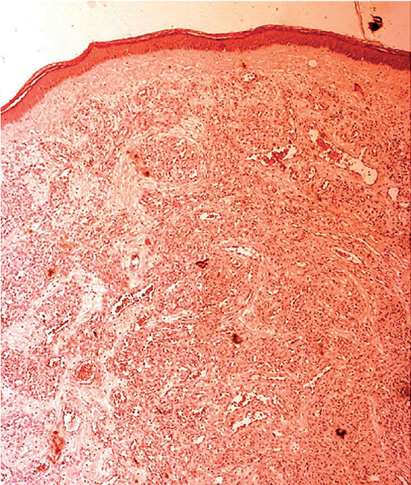 Click to show fullsize