


Department of Dermatology, Severance Hospital, Cutaneous Biology Research Institute, Yonsei University College of Medicine, 50 Yonsei-ro, Seodaemoon-Gu, Seoul 120-752, Korea. *E-mail: KYCHUNG@yuhs.ac
Accepted Aug 17, 2016; Epub ahead of print Aug 22, 2016
Primary eccrine adenocarcinoma of the skin is a rare adnexal neoplasm, with a limited number of reported cases in the literature (1). Gross features of the tumour are undefined, and variants have been reported; therefore, histological evaluation is necessary for diagnosis (1, 2). Exclusion of a visceral adenocarcinoma with cutaneous metastasis is another important criterion for confirming a diagnosis of primary eccrine adenocarcinoma (2, 3).
Traditionally, the recommended treatment for primary eccrine adenocarcinoma of the skin has been wide local excision with possible regional lymph node dissection (4); however, given the lack of published reports, there is no standard of care for management. Recently, Mohs micrographic surgery (MMS) has become an attractive treatment option for this type of tumour (3, 5, 6). For the past decade, we have treated primary eccrine adenocarcinomas of the skin based on a clinical assessment involving a total body computed tomographic and positron emission scan (positron emission tomography/computed tomography; PET/CT scan) for metastatic work-up and MMS for complete removal of the tumour. Because the role of lymph node dissection remains controversial (4), sentinel lymph node biopsy (SLNB) or regional lymph node dissection have been performed in selected cases.
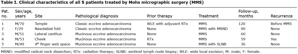
The Retrospective Clinical Data Retrieving System (Severance Hospital, Seoul, Korea) was used to obtain information from patients who were diagnosed with primary eccrine adenocarcinoma of the skin between January 2005 and November 2014. Five patients (4 males and 1 female; median age 51 years; age range 29–70 years) were identified from our search. Detailed clinical patient characteristics are summarized in Table I. On histological review, 2 patients were confirmed as having classic eccrine adenocarcinoma, and the remaining 3 patients were classified as having mucinous eccrine adenocarcinoma (Fig. S1) (5). Clinically, both types of eccrine adenocarcinoma of the skin presented with either deep or superficial nodular lesions. The tumour was confined to the head and neck area in 4 of the 5 patients, and one case of mucinous eccrine adenocarcinoma affected the finger web space. All 5 primary eccrine adenocarcinomas of the skin were treated by MMS, and various clinical features of the tumours are shown in Fig. S2. Two patients had lesions that were previously treated by either wide local excision (WLE) with adjuvant radiation (# 1) or radiation alone (# 4) and visited our clinic after initial recurrence (time to recurrence was 50 and 53 months, respectively). These 2 patients underwent MMS for control of local recurrence. The remaining 3 patients had primary occurring lesions and were initially treated by MMS.
Table I. Clinical characteristics of all 5 patients treated by Mohs micrographic surgery (MMS)
Extensive clinical examination and PET/CT scan revealed all 5 cases to be a primary eccrine adenocarcinoma of the skin. One case initially showed the presence of tumour cells in regional lymph nodes. Specifically, Patient 2 showed a mild intensity of fluorodeoxyglucose (FDG) uptake in her right neck lymph node. Modified radical neck dissection (MRND) was performed for evaluation and treatment, and metastatic involvement in 1 of 19 regional lymph nodes was confirmed. We considered SLNB in the other 4 patients with clinically and PET/CT negative node basins. Patient 5, who was diagnosed with mucinous eccrine adenocarcinoma on his finger web space, was treated by MMS along with axillary SLNB. Consistent with a negative metastatic work-up result by PET/CT scan, this patient presented with a negative SLNB specimen. Based on the unpredictable lymphatic drainage patterns of the head and neck area and the potential morbidity of neck dissection, the remaining 3 patients with lesions affecting the face were put on a biannual follow-up programme without SLNB or MRND.
Because all 5 lesions affected either the face or the finger web space, skin preservation was required. In a previous study, Wildemore et al. (7) reported that single-stage MMS with 4–5-mm margins was able to clear malignant eccrine neoplasms in all cases, suggesting that narrower margins also seem appropriate for areas, such as the face, that require maximum tissue saving. Therefore, we employed 3-mm margins for the first layer of MMS with frozen section control. Only one case of mucinous eccrine adenocarcinoma involving the lateral canthal area required 2 layers of MMS, whereas the other 4 were cleared by 1-stage MMS. Patient 2 underwent additional MRND, but no patients needed adjuvant radiation and/or chemotherapy. The mean period of follow-up after MMS was 73.0 ± 32.5 months, and only one patient presented with local recurrence during this follow-up period (# 4). This patient had undergone MMS after an initial recurrence. A second MMS was performed, and this patient has been checked every 6 months for 3 years without further recurrence or metastasis.
Primary eccrine adenocarcinoma of the skin is a rare tumour that originates in the sweat gland (1, 8). There are only about 150 cases of primary mucinous eccrine adenocarcinoma of the skin documented in the literature, and primary classic eccrine adenocarcinomas of the skin are also rarely reported (5, 9, 10). Lesions present as painless papular or nodular masses, ranging from 5 to 120 mm, with a predilection for the head and neck area (1, 2). Primary mucinous eccrine adenocarcinoma of the skin has low metastatic potential, but frequently shows local recurrence following excision (5, 6). With respect to primary classic eccrine adenocarcinoma of the skin, a high incidence of regional lymph node and visceral metastases has been reported (42% and 38%, respectively) (11); however, recently, Avraham et al. (9) reported relatively lower rates of metastasis for classic eccrine adenocarcinoma, with an overall 5-year survival rate of 81%. In the present study, one case of classic eccrine adenocarcinoma initially showed metastatic spread to a regional lymph node, but was successfully treated with MRND.
Exclusion of skin metastasis from another site is an important criterion for diagnosing and managing primary eccrine adenocarcinoma of the skin. However, given the rarity of this disease, there is little information available on the role of imaging techniques for these tumours (12, 13). While carcinoma of other organs, such as breast or gastrointestinal tract, was not detected in our patients during the follow-up period, the usefulness of supportive PET/CT scan for evaluating metastatic adenocarcinoma and establishing the diagnosis remains unclear from this small cases series. Moreover, although the histopathological results following MRND in Patient 2 and the SLNB in Patient 5 correlated with the PET/CT results, we are unable to comment on whether PET/CT is an effective technique for the nodal evaluation of this tumour. Due to the fact that a prospective large-scale longitudinal study to address these issues may be difficult for this rare tumour, we believe that PET/CT may provide benefit for the management of primary eccrine adenocarcinoma of the skin.
All 5 patients evaluated in this study are currently alive, with no evidence of metastasis after treatment with MMS. While local recurrence after MMS was observed in one case of recurrent mucinous eccrine adenocarcinoma, there was no recurrence in patients initially treated with MMS. Kamalpour et al. (10) performed a systematic review and meta-analysis of outcomes after surgical treatment of primary mucinous eccrine adenocarcinoma of the skin and found a total of 15 cases in the literature that were treated by MMS. Among these 15 patients, recurrence was observed in only 2 (13%), and there were no cases of metastasis. Adefusika et al. (5) reviewed 10 cases of sweat gland carcinomas surgically treated at the Mayo Clinic and found no episodes of recurrence or metastases after treatment with MMS. Considering the potential for aggressive behaviour following metastasis and difficult anatomical locations, such as the face and hand, MMS can be suggested as an ideal treatment option for this rare sweat gland neoplasm. Collectively, the data suggest that MMS leads to complete removal of the tumour with minimal chance for future recurrence and maximum conservation of surrounding tissue.
In conclusion, our data suggest that MMS may be an effective therapeutic option for primary eccrine adenocarcinoma of the skin, particularly in primary occurring cases. Although follow-up of 4 patients with clinically and radiologically negative node basins did not result in regional or distant metastasis, further study with a larger number of patients is needed to confirm whether in-depth investigation of the lymph nodes is necessary in this population.


 Click to show fullsize