


1Department of Dermatology, University of Occupational and Environmental Health, 1-1 Iseigaoka, Yahatanishi-ku, Kitakyushu 807-8555, Divisions of 2Dermatology, 3Clinical Pathology and 4Obstetrics and Gynecology, Japan Community Health Care Organization, Kyushu Hospital, Kyushu, and 5Department of Dermatology, Nagoya University Graduate School of Medicine, Nagoya, Japan. E-mail: natsuko-saito@med.uoeh-u.ac.jp
Accepted Aug 29, 2016; Epub ahead of print Aug 30, 2016
Impetigo herpetiformis (IH) is a rare type of generalized pustular psoriasis (GPP) occurring in pregnancy. Although patients with IH sometimes experience intrauterine growth restriction (IUGR), possibly due to lower oxygen and nutrition intake from the inflamed placenta (1), the mechanism underlying the pathogenesis of IH-associated IUGR is unknown. There have been many reports on treatment for IH-associated lower birth weight (2, 3); however, the number of case reports is insufficient to reach a consensus on IH treatment, especially when focusing on birth weight. We report here a case of IH with placental inflammation treated successfully with granulocyte and monocyte apheresis (GCAP), leading to an improvement in birth weight. We suggest that placental inflammation in IH may lead to restricted intrauterine growth, which is abrogated by GCAP.
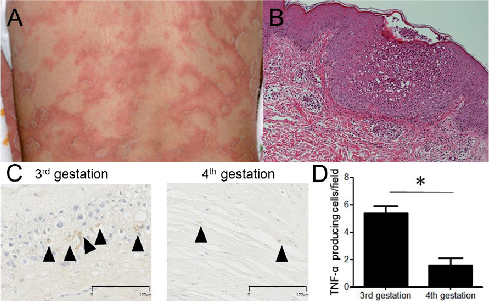
A 30-year-old woman reported circinate scaly plaques with pustules on the periphery of her trunk and her extremities at 10 weeks into her fourth pregnancy (Fig. 1A). She had experienced similar intractable skin eruptions and lower birth weight during her second and third pregnancies. Laboratory examination revealed a normal leukocyte count of 7,900/μl and a C-reactive protein level of 0.12 mg/dl (normal < 0.14 mg/dl). Skin biopsy revealed a subcorneal neutrophil-dominant infiltration, psoriasiform epidermis and perivascular infiltration of lymphocytes and a few neutrophils in the dermis (Fig. 1B). Based on the clinical course and histological examination, we diagnosed her skin eruption as an IH. She had no family history of IH or GPP. Genetic examination did not reveal any mutation in IL36RN encoding interleukin-36 receptor antagonist. She was initially treated with oral methyl-prednisolone 10 mg/day, and cyclosporine, 100 mg/day, between 10 and 16 weeks of gestation; however, her skin eruption aggravated gradually. At that time, IUGR was also observed by ultrasonographic evaluation.
Fig. 1. (A) Clinical manifestation of impetigo herpetiformis showing annular hyperkeratotic plaques with pustules on the patient’s trunk. (B) Histopathology of the skin. Haematoxylin and eosin staining of the skin showed a subcorneal neutrophil infiltration, a psoriasiform epidermis and perivascular infiltration of lymphocytes. (C) Immunostaining for tumour necrosis factor alpha (TNF-α) in placenta (arrowheads), during 3rd (left) and 4th (right) pregnancies. Scale bar: 100 μm. (D) Number of TNF-α producing cells in placenta for 3rd and 4th pregnancies. The mena (+SEM) number of TNF-α producing cells for 5 different areas was determined (original magnification ×400). *p<0.05
To relieve the intractable systemic inflammation, weekly GCAP treatment (an extracorporeal circulation therapy that removes activated granulocytes and monocytes (4)) was administered from 16 weeks gestation. The patient’s skin eruption improved after 5 GCAP treatments, and GCAP was discontinued. Two weeks after discontinuation of GCAP, however, the skin eruption gradually worsened, and then GCAP treatment was resumed. Her skin eruption improved dramatically after 5 GCAP treatments. However, 3 weeks after another cessation of GCAP treatment, her skin eruption again exacerbated. To improve her peri-operative condition, we decided to perform GCAP for her IH until 34 weeks gestation. There-fore, a total of 14 GCAP treatments were performed during her 4th gestation. After the treatment, her skin eruption improved dramatically (Psoriasis Area and Severity Index (PASI) score before and after treatment, 24.2 and 0, respectively). IUGR improved gradually and she delivered a healthy female baby with normal birth weight (1,764 g, 35 weeks’ gestation). It was speculated that GCAP might ameliorate IH-associated placental inflammation. As expected, there was less neutrophilic infiltration in the placenta after GCAP therapy following her 4th pregnancy than after her third pregnancy.
The detailed mechanism of IH-associated IUGR is largely unknown; however, the current report suggests that placental inflammation, possibly due to IH, might play a role in its pathogenesis. In fact, tumour necrosis factor α (TNF-α) producing cells, which play an important role in the pathogenesis of psoriasis, were significantly increased in the placenta during her third pregnancy with severe IUGR, compared with during her fourth pregnancy, following treatment with GCAP (Fig. 1C, D). Systemic corticosteroid is considered to be a gold standard in IH treatment (5, 6), and cyclosporine (7), narrowband ultraviolet B (UVB) (8), and anti-TNF drugs (9) are also recommended as second-choice treatments. However, the outcome of IH-associated IUGR is occasionally resistant to these treatments (10). GCAP is widely used in the treatment of inflammatory bowel diseases, but it has not yet been widely applied to skin diseases. Since GCAP suppresses TNF-α and interleukin-1β production by peripheral blood monocytes (11), the current report indicates that GCAP is useful in IH treatment, not only to improve skin eruption, but also to reduce placental inflammation and thus ameliorate IUGR. We also suggest that it is necessary to adjust treatment for patients with IH, while taking intrauterine growth into consideration. Further research into the pathogenesis of IH is needed to improve the outcome of treatment.
The authors declare no conflict of interest.


 Click to show fullsize