


1Cancer and Population Studies Group and 3Statistics Unit, QIMR Berghofer Medical Research Institute, Brisbane, Queensland, 2Institute of Cardiosvascular and Cell Sciences (Dermatology Unit), St George’s University of London, London, United Kingdom, 4Dermatology Research Centre, The University of Queensland, School of Medicine, Translational Research Institute, Brisbane, Queensland, 5Department of Dermatology, Princess Alexandra Hospital, Brisbane, Queensland, Australia, and 6CRUK Manchester Institute and Institute of Inflammation and Repair, University of Manchester, Manchester Academic Health Sciences Centre, Manchester, United Kingdom
Actinic keratoses (AKs) are common lesions that are usually diagnosed clinically. We sought to examine the accuracy of AK counts on digital photographs when compared with clinical examination counts. Skin sites of renal transplant recipients were examined clinically and on digital photographs by independent dermatologically-trained examiners. Specificity, sensitivity and Kendall’s tau-b correlation coefficient were calculated based on exact photographic AK counts as well as counts with ± 1 AK tolerance. When 138 skin sites with 305 clinical AK counts were examined for total count ± 1 AK, the sensitivity and specificity of photography was 95% and 100%, respectively. There was significant positive correlation between AK counts on photographs and clinical examination (Tb = 0.537) and correlation was even higher for total count ± 1 AK (Tb = 0.758). The results show moderate to strong concordance between AK counts on digital photographs and on clinical examination.
Key words: actinic keratosis; actinic keratosis counts; digital photographs; actinic keratosis diagnosis; virtual diagnosis.
Accepted Oct 3, 2016; Epub ahead of print Oct 4, 2016
Acta Derm Venereol 2017; 97: XX–XX.
Corr: Adele C. Green, Cancer and Population Studies Group, QIMR Berghofer Medical Research Institute, Locked Bag 2000 Royal Brisbane Hospital, Brisbane, Queensland 4029, Australia. E-mail: adele.green@qimrberghofer.edu.au
Actinic keratoses (AKs) are acquired lesions that develop as a result of chronic sun exposure (1). AKs frequently arise on white Caucasian skin and are a common presentation to dermatologists, with one study showing an estimated $920 million dollars annual spend on AK treatment in the USA alone (2).
Although histopathology can be used to diagnose AK, the high prevalence of AK, its often benign natural history, and practical and aesthetic limitations mean that in reality, the diagnosis is largely a clinical one (3, 4). AKs usually appear as red scaly papules or plaques that vary in size and shape. However, they are notoriously heterogeneous lesions and can appear hypertrophic, atrophic, pigmented or as cutaneous horns (5). The validity of the clinical diagnosis of AK has therefore often been questioned and studies have shown a positive predictive value ranging from 74–81% when compared with histopathology (6, 7). In addition, several studies have examined the reliability of measurement techniques to evaluate AKs. Weinstock et al. (8) reported poor reliability in the direct counting of AKs, although a refined technique by Atkins et al. (9), where only AKs greater than 0.5 cm were counted, showed good correlation between assessors.
The potential for using digital photography in the diagnosis of AK presents exciting possibilities such as applications in teledermatology and use in large clinical or epidemiological studies. In a small reliability study of 6 participants that compared AK counts on digital photographs with clinical counts, it was found that photographic counting was not a reliable alternative to clinical examination (10). A further study that looked at automated photographic detection of AKs based on erythema showed sensitivity of the technique ranging from 40–53% (11).
Against this background, the aim of the present study was to provide a detailed evaluation of the consistency of AK counts on digital photographs with clinical examination in renal transplant recipients, using defined areas of skin and a much larger study population than in previous studies.
The Skin Tumours in Allograft Recipients (STAR) study recruited organ transplant recipients residing in Queensland, Australia. Eligible participants were White Caucasian renal, liver or lung transplant recipients over the age of 18 years, who were at least one year post-transplantation. Details of eligibility and exclusion criteria have been described extensively elsewhere along with full details of STAR study protocol and primary outcomes (12, 13). The study in full was approved by the QIMR Berghofer Human Research Ethics Committee (project P1481) and all participants provided written consent. For the purposes of this sub-study, only renal transplant recipients were evaluated.
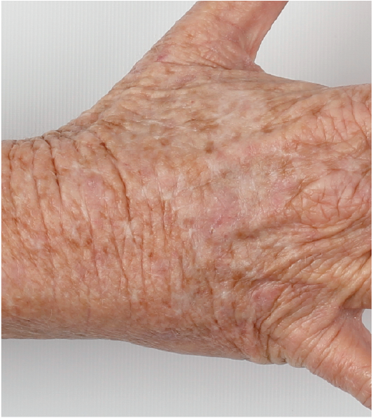
At baseline, all recruited participants underwent a full skin examination by one of several dermatologically-trained physicians. AKs were identified as erythematous papules or plaques with white to yellow scale and a consensus meeting was held at the outset of the study to establish a clear clinical definition (5). All AKs identified clinically were recorded on a body map and total numbers were summarised per skin site. In addition, high-quality photographs of the face, arms and hands (Fig. 1) of participants were taken using a Canon DS126271 digital camera (Tokyo, Japan), a Macro Lens EF-S 60 mm 1:2:8 USM, photo-studio flash lighting, 400s soft-boxes and a full-length white screen. A professional photographer was enlisted to train examiners in a standardised operating procedure and optimise images. Photographs of the face were taken front-on and in profile and the upper limbs were photographed pronated with the thumb pointed superiorly. The digital photographs of 11 defined skin sites on the face and arms (7 facial sites and 4 upper limb sites; Fig. 2) were subsequently examined by an independent dermatologically-trained physician (not one of the clinical examiners and blinded to the clinical examination outcome) who recorded total AK counts per skin site. These were then compared with clinical examination counts. The physicians ranged in experience from consultant to junior doctor and all junior physicians underwent specialised training by one of the dermatology consultants prior to commencement of skin examinations.
Fig. 1. Example of a high-quality photographic image of actinic keratoses on the dorsal aspect of the right hand.
Fig. 2. Demarcation of facial and upper limb study sites.
Concordance of AK counts on digital photographs with clinical examination was evaluated using Kendall tau-b correlation coefficient (?b). A p-value 0.05 or less was considered significant. The strength of the association was interpreted as < 0.29, weak; 0.3–0.69, moderate; 0.70–1.00, strong (14). We also examined concordance rates for total AK counts with a tolerance of ± 1 AK. Given that clinical counts can vary significantly between clinical assessors, we believe this minimal allowance of 1 AK difference would enable a fair and pragmatic assessment of the effectiveness of digital photography, particularly as tactile information is absent in photographic assessment. Sensitivity was calculated as the proportion of photographs with at least one AK identified, given at least one AK was present on clinical examination. Similarly, specificity was the proportion of photographs where no AKs were identified among the skin sites with 0 AKs on clinical examination. All analyses were performed using SPSS v21 (Armonk, NY: IBM Corp).
An unselected subset of 78 renal transplant recipients with adequate, complete, high-quality photographs was included in the present study. Multiple skin sites were examined in 28 participants, such that a total of 138 skin sites were evaluated. The mean ± SD age of included participants was 57 ± 9 years and the majority were male (67%). The mean ± SD length of time since transplantation was 9 ± 7 years. The number of AKs per skin site ranged from 0 to 14 and overall 305 AKs were diagnosed in total across all skin sites. The mean ± SD number of AKs on any single skin site was 2 ± 3.
Of 138 skin sites, 55 (40%) had the exact same number of AKs identified on digital photographs and clinical examination. When a tolerance of ± 1 AK was included, this increased to 75% (n = 104). When examining only skin sites with 5 or fewer AKs on clinical examination (n = 121), the proportion of sites with exact corresponding AK numbers identified on digital photographs was 43% and this decreased considerably (to 18%) for sites with greater than 5 AKs (n = 16). Similarly, when evaluating this result with tolerance of ± 1 AK, the percentage of skin sites that had equivalent numbers of AKs on clinical examination and digital photography fell from 70%, when restricting to skin sites with 5 or less AKs on clinical examination, to 53% when evaluating sites with > 5 AKs.
The sensitivity of detecting at least one AK on digital photographs, given at least one AK clinically, was 88% and this increased to 95% with tolerance of ± 1 AK (Table I). The specificity of digital photographs for not identifying an AK where no AK was present on clinical examination was 65%, however this was 100% when ± 1 AK was considered. There was significant positive correlation between AK counts on photographs and clinical examination (?b = 0.537). This was greater when the photographic AK counts allowed tolerance by ± 1 AK when compared with the clinical counts (?b = 0.758). The lower face (left and right combined; Fig. 2) was the skin site with the highest correlation (?b = 0.816), whilst the forearm (left and right combined) had the lowest level of correlation (?b = 0.408).
Table I. Sensitivity, specificity and Kendall tau-b correlation coefficient comparing actinic keratosis counts on digital photography against clinical examination. Without tolerance and with a tolerance of ± 1 AK for photography
Our results show that there is moderate to strong concordance of AK counts on digital photographs when compared with clinical examination and this concordance is greatly improved by allowing a tolerance of ± 1 AK. Using this allowance, the specificity of digital photography was 100% and the sensitivity of detecting at least one AK was also very high (95%). AK counts on digital photographs were highly consistent with clinical examination counts when there were 5 or fewer AKs present per skin site. This accuracy decreased notably when many (6 or more) AKs were present per skin site, thus use of this photographic technique is best applied to those with a small number of AKs.
This study is novel in analysing AK counts across a large number and variety of skin sites as well as assessing sensitivity and specificity to give an overview of the efficacy of digital photography for determining AK counts. Several studies have shown significant variation in AK counts between clinical assessors and therefore we propose that an allowance of ± 1 AK for the assessment of digital photographic counts vs. clinical counts is a progressive and pragmatic step that reflects the well-established variability in AK counts in clinical settings (8, 15, 16) and helps reconcile minor count discrepancies that have little clinical significance. Indeed, we believe that examining the effect of a tolerance of ± 1 AK in AK counts gives a better understanding of the practicality of digital photography and the potential for clinical application. Our study did not examine inter-observer reliability between photographic counts of AK which would allow an estimation of the reliability of the technique.
This study differs from our previous report by Sinnya et al. (10) which showed that digital photographs were not a reliable alternative to clinical examination for AK counts. The former study examined inter-observer reliability however, and analysis was based on mean AK counts using interclass correlation coefficients without a tolerance of ± 1 AK. Additionally, there were very few participants in that analysis (n = 6). Both studies included participants who were transplant recipients in whom correct identification of AK tends to be particularly challenging. Therefore it is possible that concordance rates may have been even higher in the general immuno-competent population.
In summary, we have shown that AK counts on digital photographs are consistent with AK counts on clinical examination, particularly in those with 5 or less AKs on a skin site. Our results show that clinical photography represents an objective documentation tool in those with limited actinic change and given the high sensitivity and specificity of the method, there is potential for use as a substitute for clinical examination where this may be necessary, such as monitoring AKs in large epidemiological studies or in remote areas where patients may have limited dermatology care.
The authors declare no conflict of interest.


 Click to show fullsize
 Click to show fullsize
 Click to show fullsize