


Department of Dermatology, No. 1 Hospital of China Medical University, 155N Nanjing Street, Shenyang 110001, China. E-mail: Pfkl2011@126.com
A 22-year-old Chinese, unmarried, woman presented to our department with pruritic annular erythematous scaly lesions on her face for 20 days. The eruptions had begun as papules and expanded progressively into annular erythematous scaly lesions. She was initially diagnosed with tinea faciei at a local clinic and was treated with topical antifungal agents for 3 weeks, but the skin lesions expanded in both size and number. No other symptoms, such as fever, malaise, headache, or arthralgia, were reported.
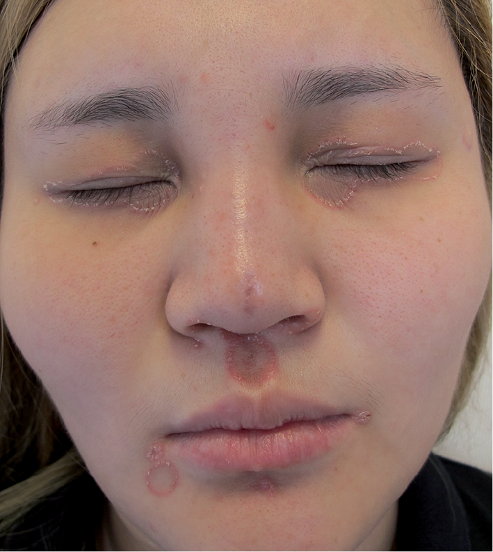
Physical examination was normal with no lymphadenectasias. Skin examination revealed multiple, annular erythematous scaly plaques with clear centres and defined borders on both eyelids, upper and lower lips and the bilateral angle of the mouth (Fig. 1). No skin lesions were observed on the mucous membranes and other skin surfaces. Laboratory tests, including complete blood counts, urine, blood sugar, and renal and liver function tests, were within normal limits. Direct microscopic examination was negative for dermatophytes. The patient refused skin biopsy for further diagnosis.
Fig. 1. Multiple, annular erythematous scaly plaques with clear centres and defined borders on both eyelids, upper and lower lips, and the bilateral angle of the mouth. Written permission has been given by the patient to publish this photograph.
What is your diagnosis?
The results of serological testing for syphilis revealed positive treponema pallidum particle agglutination assay (TPPA), positive rapid plasma reagin (RPR) (1:64), and negative HIV serotest. The patient had multiple sexual partners and had been sexually active during the past 7 years. She denied the presence of preceding chancre or any genital ulcer.
Based on the patient’s sexual contact history, clinical manifestations and the results of serological testing, a diagnosis of annular secondary syphilis was made. The patient was prescribed intramuscular benzathine penicillin (2.4 million units) once a week for 3 weeks. The skin lesions healed completely within one month and the RPR reduced to 1:4. RPR and HIV results were negative at the third and sixth month of follow-up, indicating no relapse of the disease.
The key clinical feature of this case is multiple annular erythematous scaly lesions, strongly suggesting tinea faciei. However, as the test for fungus was negative, and anti-fungal treatment did not result in any improvement, the diagnosis of tinea faciei could be excluded. Although there were no obvious manifestations of primary syphilis, taking into consideration that the skin lesion occurred after unprotected sexual contact, a diagnosis of secondary syphilis was suspected. Serological testing for syphilis confirmed the diagnosis.
Skin manifestations of secondary syphilis occur in over 80% of cases. The uncommon types of syphilides include nodular, psoriasis-like, pustular, annular, eczematous, oral mucous patches, hair loss and nail changes (1). Annular secondary syphilis is one of the uncommon forms, which often occurs in children and dark-skinned people (2). It mainly locates on the cheeks, especially close to the angle of the mouth, and forms annular, arcuate or gyrate patterns with delicate, slightly raised, infiltrated or finely scaling ridges (3). In rare situations, it is found scattered over the penis, feet, legs and nipples (4–6). This type of syphilis is rare clinically, and there is no data available about the incidence of annular secondary syphilis. Chapel (7) evaluated 105 patients with secondary syphilis, and the annular type was noted in only 6 cases.
Annular secondary syphilis should be differentiated from annular granuloma, pityriasis rosea, lichen planus, dermatophyte infection, psoriasis, erythema multiforme and drug eruption (2). Because our female patient’s skin lesions were located only on the face, clinical differential diagnoses including tinea faciei, seborrhoeic dermatitis and subacute cutaneous lupus erythematosus could be made.
Tinea faciei occurs mainly in persons who are exposed to animals with ringworm, especially cats and dogs. It is characterized by one or more circular erythematosus, slight scaling patches with distinct or indistinct borders. It can be diagnosed with direct microscopic examination (8). Seborrhoeic dermatitis appears mostly on the face, sternal region and flexures. In contrast to annular secondary syphilis, the scales in seborrhoeic dermatitis are greasy and yellowish, and itching may be severe. Serological testing for syphilis is also negative. Subacute cutaneous lupus erythematosus usually presents as scaly and polycyclic annular lesion or psoriasiform plaques, which tend to occur on the face and neck. The lesions vary from red to pink with faint violet tones, and the scale is thin and easily detached. The majority of cases of subacute cutaneous lupus erythematosus are positive for antinuclear antibody (ANA), Sjögren’s-syndrome antigen A (SSA) or B (SSB), whereas, these tests are negative in annular secondary syphilis.


 Click to show fullsize