


1Department of Dermatology, Rambam Health Care Campus and Rappaport Faculty of Medicine, Technion-Israel Institute of Technology, and 2School of Public Health, Faculty of Social Welfare and Health Sciences, University of Haifa, Haifa, Israel
Little is known about differences in epidemiological features and prognosis between pemphigus vulgaris (PV) and pemphigus foliaceus (PF). The objective of this study was to compare PV and PF patients regarding ethnic variations and mortality rates. Mortality of PV and PF patients was compared with age- and sex-matched control subjects in the general population. The study cohort comprised 207 patients with PV and 30 with PF diagnosed during the period 2000 to 2015. The incidence rate of PV among Jews was 3.6-fold high-er than among Arabs (p < 0.001), whereas no ethnic predisposition to PF was noted (p = 0.379). The risk of death for patients with PV was almost 3-fold higher than in the general population (standardized mortality ratio (SMR) 2.6). For patients with PF, the risk of mortality was not significantly increased relative to the general population (SMR 1.4). There is a racial predisposition to PV, whereas PF is sporadic. Mortality among patients with PV is higher compared with PF and the general population.
Key words: pemphigus; vulgaris; foliaceus; epidemiology; mortality; prognosis.
Accepted May 22, 2017; Epub ahead of print May 24, 2017
Acta Derm Venereol 2017; 97: xx–xx.
Corr: Khalaf Kridin, Department of Dermatology, Rambam Health Care Campus, POB 9602, Haifa 31096, Israel. E-mail: dr_kridin@hotmail.com
Pemphigus is a rare, chronic, potentially life-threatening, autoimmune blistering disease of the skin and mucous membranes. It has 2 major subtypes: pemphigus vulgaris (PV) and pemphigus foliaceus (PF). The aetiopathogenesis of pemphigus is characterized by acantholysis and intraepidermal blister formation, resulting from IgG autoantibodies directed against desmoglein (Dsg) 3 (PV) and/or Dsg 1 (PF), 2 transmembrane desmosomal glycoproteins (1, 2).
Pemphigus has an uneven geographical and ethnic distribution. We recently reported that the incidence rate of all variants of pemphigus in northern Israel was 3-fold higher among Jews than among Arabs (the major ethnic populations in Israel), with an overall annual incidence of 7.2/million (3). The annual incidence of PV is variable, and ranges from 0.76/million in Finland (4) to 16.1/million in Jerusalem (5). In most populations PF is less common than PV, and the estimated annual incidence varies between 0.5–1.0/million in Western Europe (6) to 6.7/million in Tunisia (7). PF is rare and sporadic worldwide, and its ethnic predominance is yet to be demonstrated (8).
The mainstay treatments for pemphigus are systemic corticosteroids and immunosuppressive therapy. The prognosis for patients with pemphigus has improved greatly since the introduction of corticosteroid therapy; nevertheless, pemphigus remains a potentially life-threat-ening disease (6), with a relatively high mortality rate after diagnosis, ranging from 5% to 30% during various lengths of follow-up (9–11). In 2 recent studies from the UK and Taiwan, the risk of death was calculated as 2-to-3-fold higher than in the control or general population (12, 13). We have demonstrated previously that overall mortality of patients with pemphigus is 2.4-times greater than for the general population (14).
The differences in the risk of overall mortality in patients with PV and PF relative to the general population have not yet been investigated. Moreover, the mortality of PF, as a distinct disease relative to the general population, has not been profiled.
The objectives of this study were to investigate and compare the 2 major subtypes of pemphigus regarding: (i) the incidence rate in 2 distinct ethnic populations; and (ii) mortality rates compared with the general population, by tracking an immunopathologically validated large cohort of consecutive PV and PF patients over an extended period of time. The data obtained represent the first comparison between PV and PF patients regarding mortality rates relative to the general population.
The study comprised all Israeli patients in Haifa (population: 875,000) and the northern (population: 691,000) districts with a new diagnosis of PV or PF from the beginning of January 1990 to the end of December 2015 (2008 census). The study cohort also included 16 patients diagnosed between 1985 and 1989, and followed up intensely during the study period.
To assess the incidence of PV and PF for an extended duration, we conducted a retrospective cohort study from January 2000 to December 2015. Incidence data are more reliable from 2000 due to the installation of a computerized system in our institute, which enables confirmation of every PV or PF case, and due to a better accessibility to the Israeli census. Rambam Health Care Campus is the tertiary referral centre providing dermatology services for the entire north-west region of Israel, and is the only hospital in the region that offers diagnostic laboratory immunopathology sample analysis. Thus, regional patients with suspected pemphigus are referred to our centre. All the patients included in the incidence analysis were Israeli citizens living in one of the two districts.
Inclusion criteria for patients with PV were: (i) presence of skin blisters and/or erosions on mucous membranes; (ii) suprabasal intraepidermal acantholysis on histopathological examination of skin and/or mucosa; (iii) intraepidermal intercellular IgG and/or C3 deposits by direct immunofluorescence (DIF); or intercellular circulating antibodies demonstrated by using monkey oesophagus and a standard indirect immunofluorescence (IIF) technique, or the presence of anti-Dsg 3 ± anti-Dsg 1 autoantibodies, measured by enzyme-linked immunosorbent assay (ELISA) or immunoblotting (15).
Inclusion criteria for patients with PF were: (i) presence of skin blisters or erosions; (ii) lack of mucosal lesions; (iii) intraepidermal acantholysis compatible with PF on histopathological examination; and (iv) intraepidermal intercellular IgG and/or C3 deposits by DIF, intercellular circulating antibodies demonstrated by a standard IIF technique; or presence of anti-Dsg 1 autoantibodies, with lack of anti-Dsg 3 autoantibodies, measured by ELISA or immunoblotting (15). Patients diagnosed with subtypes other than PV or PF were not included in our study.
Cases were identified using the hospital’s computerized database for International Statistical Classification of Diseases and Related Health Problems – 9th Revision (ICD-9) diagnosis of pemphigus. Before inclusion, 2 of the authors (KK, SZ) verified that selected cases fulfilled all the diagnostic criteria for pemphigus and reviewed medical files in order to exclude prevalent cases for the incidence analysis.
The mainstay of therapy in our department for both PV and PF has been approximately 1.5 mg/kg/day oral prednisone. Adjuvants, such as oral azathioprine, methotrexate or cyclophosphamide, were added when prednisone failed to control the disease or when relapses occurred. Rituximab, plasmapheresis, and intravenous immunoglobulin were administered to relatively small numbers of patients (n = 25), due to severe or recalcitrant disease refractory to the above-mentioned treatments. When in complete remission, all treatments were gradually tapered and withdrawn, but re-administered when relapses occurred and then usually continued indefinitely at the lowest possible dose.
To evaluate the mortality of PV and PF patients, we used data from a prospective cohort of consecutive PV and PF patients who were actively followed for a minimum of 12 months, or until death (if it occurred during the first year following diagnosis). Patient date of death was ascertained by linking the study cohort with the National Registry of Deaths Database of Haifa district. Patients not listed in that database were considered survivors. All patients were followed up from the onset of pemphigus until 29 December 2016 (for at least 12 months from diagnosis), or until death. The study had approval from the institutional Ethics Committee of our medical centre.
Incidence rates were estimated as ratios of the number of newly diagnosed cases of pemphigus for the years 2000 to 2015 divided by 16 times the 2008 census population, obtained from the Israel Central Bureau of Statistics (www.cbs.gov.il). Continuous variables are presented as means ± standard deviation (SD), while categorical variables are presented as percentages. For each incidence rate, an exact 95% confidence interval (CI) was obtained, based on the Poisson distribution. Incidence rates were estimated for the whole population and separately by age category (< 45, 45–65, ≥ 65 years), sex, geographical region and ethnic group. Crude comparisons between incidence rates relied on exact Poisson inference. Comparisons in continuous variables between different age categories and ethnic groups were performed using Students t-tests for independent samples.
Observed survival curve from the onset of PV and PF patients was estimated using the Kaplan–Meier method. The expected survival curve of the study cohort was computed according to Hakulinen’s method (16), using sex-, age- (1-year classes), and calendar year-specific (1-year classes) mortality rates for the population of Israel (Israel Central Bureau of Statistics). To compare the observed and expected survival rates, we calculated standardized mortality ratios (SMRs), the ratio of the observed to the expected number of deaths, with 95% Poisson confidence intervals. The expected number of deaths was calculated by multiplying person-years of each sex-, age (1-year classes)-, and calendar year-specific stratum of the study cohort by the corresponding mortality rate of Israel population, summed up across all strata. All analyses were performed using STATA statistical software version 8.2 (StataCorp, College Station, TX, USA).
The study cohort comprised 237 consecutive patients with PV and PF. In total, 207 (87.3%) patients met the diagnostic criteria of PV and 30 (12.7%) those of PF.
Pemphigus vulgaris. A total of 75 (36.2%) patients were male and 132 (36.8%) female; a male-to-female ratio of 1:1.8. Mean ± SD age at diagnosis was 52.9 ± 16.0 years (range 0–90.0, median 53.0 years). Men were significantly younger than women (49.3 ± 16.2 vs. 54.9 ± 15.6 years, respectively, p = 0.01), and Arabs were younger than Jews (44.3 ± 15.4 vs. 54.5 ± 15.6 years, respectively, p = 0.001) (Table I).
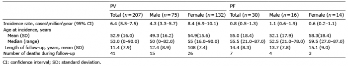
Table I. Characteristics of 237 patients with pemphigus vulgaris (PV) and pemphigus foliaceus (PF) followed-up from January 1990 to December 2015
The majority of patients were of Ashkenazi Jewish origin (62.8%), followed by Sephardic Jews (20.8%) and Arabs (15.5%). In 2 patients, the ethnic descent could not be determined.
Pemphigus foliaceus. Altogether, 16 (53.3%) males and 14 (46.7%) female patients were included in this subgroup, representing a male to female ratio of 1.1:1.
The mean ± SD age at diagnosis was 55.0 ± 18.4 years (range 21.0–87.0, median 55.5). Men were younger than women (52.1 ± 17.9 vs. 58.3 ± 18.4 years, respectively, p = 0.35), and Arabs were younger than Jews (48.6 ± 16.2 vs. 57.3 ± 18.5 years, respectively, p = 0.28) (Table I).
The ethnic distribution of the patients was: 43.3% Ashkenazi Jews, 30.3% Sephardic Jews and 26.7% Arabs.
The incidence rates of the 2 subtypes were estimated during the years 2000 to 2015.
Pemphigus vulgaris. The overall annual incidence of PV was 6.4/million (95% CI 5.5–7.5). The incidence of PV increased sharply with age and was significantly higher amongst women than men (8.4 vs. 4.3/million/year, respectively, p < 0.0001).
Interestingly, the incidence rate in the Jewish population was almost 3.5-fold higher than that in the Arab population (8.8 vs. 2.5/million/year, respectively, p < 0.0001). When age-standardized incidence rates were calculated using the European standard population as a reference, the discrepancy between the 2 populations persisted (8.6 vs. 3.5/million/year, p < 0.0001).
Pemphigus foliaceus. The mean annual incidence of PF was 0.8/million (95% CI, 0.5–1.3). Calculated age-specific rates revealed a clear and marked increase in incidence of PF with age. The incidence was higher in men (1.1/million/year) than in women (0.6/ million/ year), although lacking a statistical significance (p = 0.123). A slight predominance of males persisted in all age categories.
In contrast to PV, no significant ethnic predisposition was registered; the incidence rate among Jews was slightly higher than among Arabs (1.0 vs. 0.7 cases/million/year), albeit not statistically significant (p = 0.379). Adjustment for age using the European standard population as a reference reinforced the lack of statistical difference (0.9 vs. 1.1 cases/million/year in Jews and Arabs, respectively; p = 0.451).
Pemphigus vulgaris. Overall, 207 patients were followed for at least 12 months or until death (if it occurred earlier), contributing 2,349.7 person years. The mean ± SD length of follow-up was 11.4 ± 7.9 years.
A total of 41/201 patients (19.8%) died during the follow-up period, of whom, 10/41 (24.4%) died within one year of being diagnosed with PV (overall 1-year mortality rate was 4.8% [10/27]. Patients who survived less than one year were significantly older at the time of diagnosis than those who survived at least one year but died later (76.2 ± 8.5 vs. 60.0 ± 12.2 years, respectively, p = 0.0004). Furthermore, 46.3% of those who died during the study period died within 10 years of initial presentation. Patients who survived less than 10 years were significantly older at the time of diagnosis than those who survived at least 10 years and died later (9.8 ± 11.5 vs. 58.9 ± 12.8 years, respectively, p = 0.0068).
The 1-, 5-, 10-, 15- and 20-year overall survival rates were 95.2%, 92.9%, 89.5%, 80.2% and 65.3%, respectively, in the entire population of patients with PV (Table II). No significant differences in survival rates were noted between sexes (p = 0.57). The median overall survival period amongst patients who died was 10.1 years (range 0.2–29.8 years).
Table II. Estimated overall survival following the diagnosis of pemphigus vulgaris (PV) and pemphigus foliaceus (PF)
The survival rates for patients with PV were strikingly lower compared with the general population. Kaplan–Meier survival curves for PV cohort were lower than expected in the age- and sex-matched general populations; in particular as the length of follow-up increases (Fig. 1). Overall, patients with PV experienced 2.6-fold the expected number of deaths (SMR=2.6; 95% CI, 1.9–3.6).
Fig. 1. Survival among patients with pemphigus vulgaris (PV) and pemphigus foliaceus (PF) diagnosed until December 2015, compared with expected survival in age- and sex-matched population.
In sex-specific analysis, the SMRs for patients with PV were significantly higher for both men (SMR, 2.8; 95% CI 1.6–4.6) and women (SMR, 2.6; 95% CI 1.7–3.7) (Table III).
Table III. Observed and expected deaths and standard mortality ratios in patients with pemphigus vulgaris and pemphigus foliaceus stratified by sex and age at incidence
Pemphigus foliaceus. A total of 30 patients were followed for at least 12 months, contributing 433.1 person-years. The mean ± SD length of follow-up was 14.4 ± 8.3 years. Seven of the 30 (23.3%) patients diagnosed in the above period died. None died within the first year, 1 died within 5 years and 2 within 10 years of their initial diagnosis of PF. Patients who survived less than 10 years were older at the time of diagnosis than those who survived at least 10 years and died later, without reaching the level of statistical significance (75.5 ± 14.9 vs. 57.8 ± 13.5 years, respectively, p = 0.819).
The 1-, 5-, 10-, 15- and 20-year overall survival rates were 100%, 96.7%, 92.5%, 81.2% and 81.2%, respectively, in the population of patients with PF (Table II). The median overall survival of patients who died was 14.0 years (range 1.9–35.5 years). No significant sex-specific differences in the survival were observed.
Relative to expected age and sex-specific overall death rates in the general population in Israel, there was a greater than 1.4-fold excess of mortality among PF patients, with a SMR of 1.4 (95% CI 0.6–2.9) for the population of patients with PF, albeit not statistically significant. The excess mortality was slightly higher in men than in women, with a SMR of 1.7 (95% CI 0.5–4.5) among men and 1.1 (95% CI 0.2–3.1) among women (Table III).
This population-based study demonstrated that the incidence rate of PV in northern Israel estimated at 6.4/million/year, is amongst the highest reported worldwide (17), whereas the incidence of PF is comparable to that in western countries (6).
In this analysis, we found a 3.5-fold higher incidence rate of PV in the Jewish population compared with the Arab population, while the incidence of PF was not related to ethnicity. These results are in line with fact that a genetic susceptibility associated with the HLA types has been well documented in Ashkenazi Jewish patients with PV. Previous studies demonstrated that HLA-DRB1*04 and HLA-A*10 was markedly increased among PV patients belonging to this population (8, 18). Recently, a polymor-phic variant in ST18 encoding a pro-apoptotic molecule was found to be significantly associated with PV in Jewish and Egyptian cohorts, but not in a German sample (19). Different access to healthcare services cannot account for these ethnic variations because the 2 populations share the same healthcare system. Non-endemic PF seems to be sporadic without any reported ethnic predisposition. Three foci of endemic PF were described; (i) Fogo selvage (or endemic PF), which occurs in some subtropical areas of Brazil, with data being extensively collected in the Limao Verde Amerindian reservation in the state of Mato Grosso do Sul. The prevalence of the disease was estimated at 3–5% in this population (20); (ii) Columbian PF, which has been reported in the El Bagre area in Northern Colombia, where the prevalence of the disease is close to 5% (21); (iii) Tunisian PF, which mainly occurs in the southern area of Tunisia, Algeria, Morocco as well as in some sub-Saharan countries, such as Mali (7, 22).
The higher incidence rate of PV observed in woman than in men, and the 1:1.8 male-to-female ratio in the PV group, are consistent with known female preponderance of pemphigus (17). On the other hand, we observed a slightly higher incidence rate of PF among males compared with females, albeit insignificant. These findings are in contrast to the demographic characteristics of a Tunisian PF cohort, where a male-to-female ratio of 1:4.1 was registered, with a strikingly higher incidence rate among young women (7). Notwithstanding, the majority of patients with endemic PF in northern Colombia are men (95%) (21). The epidemiology of PF may be related more to environmental factors than to ethnicity (20, 23).
The current study demonstrated that survival in patients diagnosed with PV was significantly lower than that expected in the general population. Overall, patients with PV had a 2.6-fold increase in mortality compared with the general population. The risk for death in patients with PF was 1.4-fold higher than the general population, without reaching statistical significance (95% CI, 0.6–2.9). To the best of our knowledge, the current study is the first to report that mortality in patients with PF is comparable to age- and sex-matched control subjects. Compared with their PV counterparts, patients with PF showed consistently higher survival rates at 1-, 5-, 10- and 20-years of follow-up, and the median overall survival among PF patients who eventually died was longer (14.0 vs.10.1 years, p = 0.05, respectively). The higher mortality rate observed in the PV cohort was consistent with the fact that it is a more life-threatening disease, characterized by a high mortality rate (24), while PF has a more benign and chronic course, which tends to persist for months to years.
Pemphigus is associated with a relatively high mortality rate after diagnosis, ranging in the literature from 5% to 30% during various lengths of follow-up (9–11, 25–28). Of note, the vast majority of the survival data reported among pemphigus patients did not differentiate between the different variants of the disease as distinct entities. Moreover, mortality among patients with PF relative to control subjects was not investigated in the past. Our study contributes an epidemiological figure of all-cause mortality for PF patients relative to the general population, which was not found to be significantly increased.
This study has some limitations. First, due to the fact that it was held in a tertiary healthcare centre, we may have missed mild cases of pemphigus managed by the community dermatologist. In our region, however, community dermatologists are highly unlikely to manage patients with pemphigus without referring to a hospital-based department of dermatology. Secondly, our study provides data from a relatively small region populated with up to 1.6 million inhabitants. Another limitation is the theoretical probability of missing death cases in patients who moved outside the country or changed their names, and thus would not appear in the Israeli death registry.
PV was 7-fold more frequent than PF in the study population. PV was found to be strikingly more frequent among Jews, whereas no ethnic predisposition was registered in the incidence of PF. There is a marked decrease in the long-term overall survival in patients with PV relative to their PF counterparts and the general population. Patients with PF do not seem to have significantly increased mortality compared with the age- and sex-matched general population.
The authors declare no conflicts of interest.


 Click to show fullsize
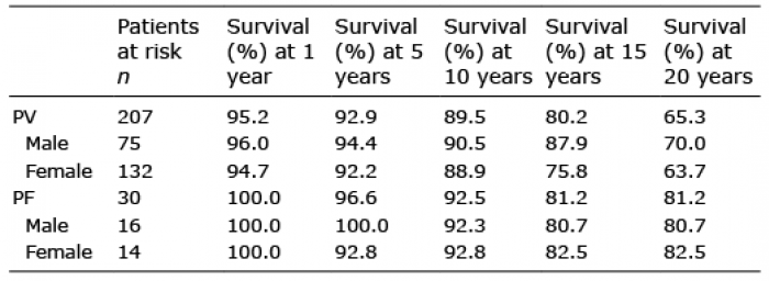 Click to show fullsize
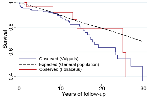 Click to show fullsize
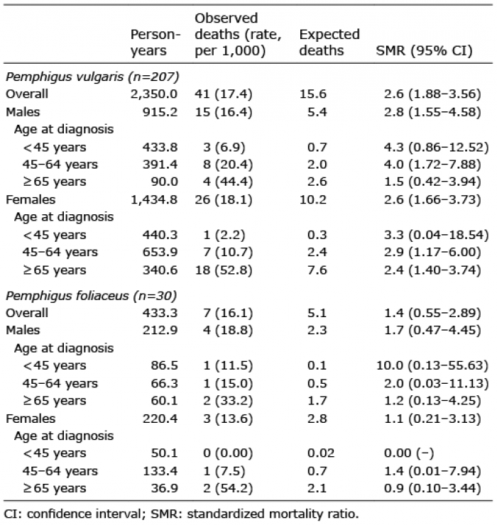 Click to show fullsize