


1Department of Pediatrics, OLV Hospital Aalst, BE-9300 Aalst, 2University of Louvain, Louvain, Belgium and 3Department of Dermatology, University Medical Center Schleswig-Holstein, Campus Kiel, Germany. *E-mail: dirk.van.gysel@olvz-aalst.be
#These authors contributed equally to this manuscript.
Accepted Oct 19, 2017; Epub ahead of print Oct 23, 2017
The incidence of measles has reduced since the availability of live attenuated vaccines. However, despite all the efforts made to eradicate measles, outbreaks still occur in Europe (1–3). The persistence of measles can be explained by importation of the virus from other countries, vaccination refusal and, less frequently, primary vaccination failure. Furthermore, waning measles-specific IgG titres after vaccination and lack of natural immunological boosting due to increasing vaccination rates lead to secondary vaccination failure. Thus, measles can occur even in fully vaccinated individuals when exposed to wild-type measles in an outbreak setting (1, 4, 5). We report here a case of a 15-year-old boy who developed measles despite having had 2 previous doses of live attenuated measles-mumps-rubella (MMR) vaccine, administered at the ages recommended by the WHO (6). The aim of this publication is to remind healthcare workers of the increasing occurrence of measles in previously vaccinated individuals, so called “modified” or “secondary” measles. Due to the possible life-threatening complications of measles, and in order to prevent further spread of the disease, healthcare workers should be familiar with the clinical course of (re)-infection and altered laboratory findings after vaccination (7).
A 15-year-old boy was referred to the emergency department with high fever and a generalized rash. His symptoms had started 3 days earlier with a strong headache, general malaise, conjunctivitis and high fever (up to 39.9°C). On day 2 a rash appeared, starting on his face and spreading to his neck, upper and lower trunk and extremities. Approximately 10 days before the onset of symptoms, the patient had visited Wallonia, a part of Belgium recently affected by an outbreak of measles. The patient had been vaccinated twice for measles: at the age of 2 years and at the age of 10 years. He had no history of immunodeficiency or immunosuppression. His current medication consisted of methylphenidate hydrochloride (Equasym®) 30 mg/day for ADHD.
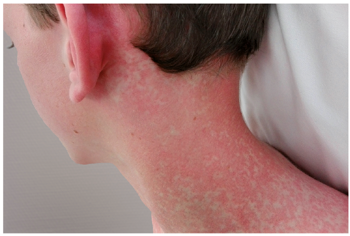
Physical examination revealed that his general condition was impaired because of the symptoms mentioned above, with an erythematous, confluent, maculopapular rash (Fig. 1) over his entire body except for the palms, soles and head. His body temperature was 38°C 2 h after intake of paracetamol. Neurological examination revealed terminal nuchal rigidity. Kernig’s and Brudzinski’s tests were negative. No other abnormalities were observed on general physical examination.
Fig. 1. Morbilliform rash in the neck on admittance to hospital (day 3 of the exanthem).
Laboratory investigations documented a white blood cell count of 5,670/µl (normal range 4,000–10,000/µl) and a C-reactive protein (CRP) of 51.8 mg/l (normal <5.0 mg/l). IgM and IgG titres for measles were 0 and >300 U/l respectively. IgM titres for both rubella and Epstein-Barr virus (EBV) were negative, whereas IgG titres were positive. Lumbar puncture excluded meningitis. To differentiate from other infectious agents, a standard set of 20 PCRs was performed on a nasopharyngeal aspirate and was found to be negative. Because of the very suggestive clinical features and course a second measles-specific RNA-PCR analysis on a saliva sample was requested, and proved to be positive.
Because of the risk of secondary bacterial infection treatment with ceftriaxone 2×2 g IV was initiated. Over the next 3 days the patient underwent clinical improvement and his fever gradually diminished. The erythematous morbilliform exanthem started to fade on the trunk and turned into a purpuric rash on the legs. On day 3 of hospitalization, ceftriaxone was stopped as all blood cultures were sterile and the patient was discharged from the hospital in good general health apart from a mild headache and low-grade fever.
Modified measles often has a milder, less characteristic, clinical course compared with primary measles and can therefore easily be misdiagnosed (1, 7–9). This is illustrated by Rota et al. (8), who documented 2 healthcare workers with modified measles due to contact with patients with a primary infection. One healthcare worker presented with a rash starting on the abdomen and spreading to the neck, unlike the classical craniocaudal spread. The other healthcare worker presented with a prodromal phase with fever and headache, without coryza, conjunctivitis or cough. The current case presented with all the symptoms of classical measles (e.g. prodromal phase with general illness, coryza, cough, followed by the characteristic maculopapular rash spreading craniocaudally to the trunk and extremities) but in an attenuated form. Symptoms can thus be modified, but might as well be very similar to primary infection with measles. In all cases however, symptoms are milder and resolve faster. Furthermore, previous studies suggest that secondary infection is less contagious than primary measles (8). Due to its atypical clinical findings and course it can be difficult to differentiate modified measles from other conditions with a maculopapular rash, such as other (viral) infections or adverse drug reactions (10).
In order to diagnose measles unquestionably, laboratory confirmation is needed. Detection of virus-specific IgM antibodies is the standard laboratory method used to diagnose acute measles infection. However, IgM might be false-positive or false-negative and the IgM response might be short-lived or even absent in previously vaccinated individuals (4, 11). When IgM is absent, but measles is clinically suspected, measurements of IgG titres and/or IgG-avidity can help in the diagnosis of acute measles. A 4-fold increase in IgG antibodies between 2 paired samples, collected in the acute phase and the convalescent phase, respectively, indicates acute measles infection and might be present even in the absence of IgM titres (11, 12). Analysis of IgG avidity distinguish-es primary infection, characterized by low-avidity IgG antibodies, from re-infection cases in individuals with previous immunological response due to vaccination or natural immunization 4 months before onset of symptoms, characterized by high-avidity IgG antibodies (11, 13). It is clear that serological results are often inconclusive. Therefore, RNA-PCR testing should be performed using nose and throat swabs in conjunction with serological testing (11, 12). However, restricted viral replication in previously immune patients may lead to false-negative results (3, 11, 13). The sensitivity of the test also depends on the timing of sample collection and transport conditions. Sampling for viral PCR should preferentially be performed within the first 3 days after rash onset, whereas blood samples for IgM titres should be collected one week after rash onset (3).
In conclusion, healthcare professionals should be aware of the occurrence and diagnostic challenge, both clinically and in the laboratory, of modified measles. Serological testing in combination with PCR analysis on nose or throat swabs is necessary to diagnose modified measles.


 Click to show fullsize