


1Institute of Dermatology, Chinese Academy of Medical Sciences and Peking Union Medical College, Nanjing, Jiangsu, 210042, and 2Jiangsu Key Laboratory of Molecular Biology for Skin Diseases and STIs, Nanjing, China. E-mail: whs33@vip.sina.com
#These authors contributed equally to this work.
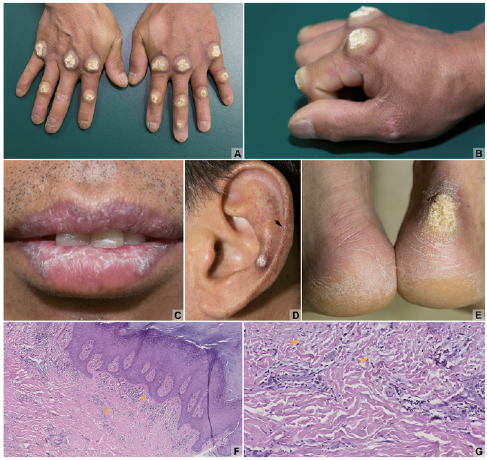
A 50-year-old man presented with hyperkeratotic scales on his lips, asymptomatic, round, discrete, hyperkeratotic, verrucous nodules on the dorsa of the interphalangeal and metacarpophalangeal joints, the left ear, right heel (Fig. 1A–E), and poikiloderma over his fingers and left ear (Fig. 1B). The lesions had gradually increased over a period of 20 years. Laboratory examinations revealed reduced platelet number (92×109/l), positive antinuclear antibodies (1:160, speckled pattern), anti-dsDNA and anti-SSA/Ro. Histopatho-logy of biopsied foot lesions revealed marked hyperkeratosis, acanthosis and hypergranulosis (Fig. 1F). There was lympho-cyte infiltration around the vessels and in the upper dermis, and mucin deposition in the superficial and mid-dermis (Fig. 1G). Direct immuno?uorescence of IgG and complement 3 was negative. After treatment with methylprednisolone, 8 mg q.d., hydroxychloroquine 100 mg and viaminate 50 mg b.i.d., topical 0.05% halometasone cream b.i.d. for 1 month, the patient reported that most of the lesions became flatter.
What is your diagnosis? See next page for answer.
Fig. 1. (A, B, E) Verrucous hyperplastic plaques and nodules on the interphalangeal and metacarpophalangeal joints, and right heel. (C, D) Hyperplastic plaques on the lips, nodule and the poikiloderma (arrow) on the left ear. (F, G) Histopathological aspects of the right heel lesion, demonstrating lymphocyte infiltration (arrowhead) and mucin deposition in the dermis (arrow), original magnification: (F) ×50, (G) ×200.
Acta Derm Venereol 2018; XX: XX–XX.
Diagnosis: Hypertrophic lupus erythematosus
Hypertrophic lupus erythematosus (HLE) is a variant of
chronic cutaneous lupus erythematosus (CLE). HLE with lips or oral mucosa lesions is seldom mentioned in the literature (1). The similarity with cutaneous lesions has been reported previously and it seems to arise via the same mechanisms (1, 2). The primary histological change is marked hyperkeratosis hyperplasia associated with features of classic CLE (3). Although the manifestation of marked hyperkeratosis is characteristic and helpful for diagnosis of HLE, the patient could not be conclusively interpreted as having lupus via pathology. Histopathology of a foot lesion revealed no signs of keratinocytic atypia, differentiating it from squamous cell carcinoma. The patient’s clinical manifestations closely resemble hypertrophic lichen planus and several other hyperkeratotic conditions, such as knuckle-pads, and warts on the hands and callus on the heel. However the immunological disorder and the good effect of hydroxychloroquine suggest HLE. This diagnosis was made principally based on the combination of distinctive hypertrophic lesions, reduced platelet counts, positive antinuclear antibody and anti-dsDNA, which fulfilled the criteria for systemic lupus erythematosus (SLE) according to the 1997 American College of Rheumatology classification criteria and 2012 Systemic Lupus International Collaborating Clinics criteria (4). CLE can be the first sign of SLE, which appears in 25% of patients with SLE, while up to 45.7% of discoid lupus erythematosus (DLE) precedes SLE (5, 6). Widespread DLE lesions below the neck, anaemia, leucopaenia, positive antinuclear antibodies and anti-dsDNA antibodies may be positive predictors for progression to SLE (6). Unhealed lesions of CLE may also transform to squamous cell carcinoma after some years (7). Taking all these factors into consideration, overall assessment of clinical material and systemic examinations, and long-term follow-up are important for patients with DLE.
HLE are often chronic and refractory to drugs. Although hydroxychloroquine has been proven to have good efficacy and safety in the treatment of HLE, fulminant hepatic failure had also been reported soon after the start of treatment (8–10). The current case was treated effectively with a combination of hydroxychloroquine, low-dose corticosteroids and viaminate for 1 month. Viaminate is a derivative of retinoid, which was prepared from all-trans retinoid acid and p-amino-benzoic acid ethyl ester (11). Clinically, viaminate appears to possess the therapeutic effects of, for example, acitretin, but with less toxic side-effects (11, 12). However, clinical trials are needed to assess the full effects of this combination in treatment of HLE.
This manuscript was supported by grants from the Chinese Academy of Medical Sciences Innovation Fund for Medical Sciences (CIFMS-2016-I2M-1-005), National Natural Science Foundation of China (81371751), and the Natural Science Foundation of Jiangsu Province of China (BK20141065).


 Click to show fullsize