


1Institute for Environmental and Gender Specific Medicine, Juntendo University Graduate School of Medicine, 2-1-1 Tomioka, Urayasu, Chiba 279-0021, 2R&D laboratories, Pola Pharma Inc., 560 Kashio-cho, Totsuka-ku, Yokohama, Kanagawa, 3Atopy (Allergy) Research Center, Juntendo University Graduate School of Medicine, Tokyo 113-8421, and 4Department of Dermatology, Juntendo University Urayasu Hospital, Chiba, Japan. *E-mail: ktakamor@juntendo.ac.jp
#These authors contributed equally to this work.
Accepted Jun 26, 2018; Epub ahead of print Jun 29, 2018
Atopic dermatitis (AD) is characterized by chronic cutaneous inflammation and dryness with skin barrier dysfunction. Chronic and intractable itch in patients with AD markedly diminishes their quality of life (1, 2). Low-threshold mechanical stimuli, such as wool fibres, are irritants to the skin of patients with AD (3). This phenomenon is known as alloknesis (touch-evoked itch) (4), and the hypersensitivity results in an itch-scratch cycle, which causes refractory AD (5). The number of epidermal nerve fibres in patients with AD is higher than in healthy individuals, and a reduction in intraepidermal nerve fibres by psoralen and ultraviolet A (PUVA) therapy improves both pruritus and dermatitis scores in patients with AD (6). Topical treatment of the nerve repulsion factor semaphorin 3A significantly reduced intraepidermal nerve density, which suppressed scratching behaviour and improved dermatitis scores in AD model NC/Nga mice induced by Dermatophagoides farina body ointment (7). Therefore, intraepidermal nerve fibres are a likely pathogenic factor for itch hypersensitivity, including alloknesis in AD (8), but conventional treatments for alloknesis are limited. Therefore, it is necessary to develop preventative and therapeutic approaches for alloknesis in AD.
Film dressings are transparent sheets coated with an adhesive, and some are thin enough (several 10 µm) to be almost invisible on the skin and do not interfere with tactile transmission. They are used for wound treatment to provide an appropriately moist environment and act as a barrier to contamination. However, it is unclear whether film dressings prevent epidermal hyper-innervation or alloknesis in dry skin-based diseases, such as AD. This study evaluated 2 different thicknesses of film dressings; Tegaderm™ (TDM, 24 µm; 3M Japan, Tokyo, Japan) and Perme Roll™ Lite (PMR, 8 µm; Nitto, Tokyo, Japan), using 2 murine dry skin models for evaluation of epidermal hyper-innervation or alloknesis efficacies.
All animals were housed in the experimental animal facility of Juntendo University Graduate School of Medicine, and all animal procedures were approved by the Animal Care and Use Committee of Juntendo University Graduate School of Medicine. The study conformed to the National Institute for Health guidelines for animal research.
The protocol for induction of epidermal hyper-innervation of male ICR mice (10 weeks old; SLC Japan, Shizuoka, Japan) by cutaneous barrier disruption has been described previously (9). A 20-mm square of TDM, PMR, or nothing (positive control) was applied to an acetone-treated area immediately after 5 min of acetone treatment, and distilled water-treated mice were used as a negative control (n = 6 per group). Skin samples were taken after an additional 48 h, and the nerve fibres in 20-µm cryosections were stained immunohistochemically using rabbit anti-protein gene product 9.5 (PGP9.5, 1:400 dilution; Enzo Life Sciences Inc., NY, USA).
The protocol for the alloknesis assay by cutaneous barrier disruption has been described previously (4). Briefly, a cotton soaked with a mixture of acetone and diethylether (1:1) was applied to the rostral section of the shaved back of C57BL/6NCrSlc mice (10 weeks old; SLC Japan, Shizuoka, Japan) for 15 s, followed immediately by applying distilled water for 30 s (AEW treatment), and the negative control group was treated only with distilled water for 45 s, twice daily for 8 days. On the day after the last AEW treatment, 5 separate innocuous mechanical stimuli were applied using a von Frey filament (0.7 mN; DanMic Global, CA, USA) to the AEW- or water-treated site to determine the sections suitable for alloknesis assessment (AEW; n = 12, water; n = 4). Next, we applied TDM, PMR, or nothing (positive control) with a diameter of 8 mm on the right or left rostral section (alloknesis spot) of the AEW-treated site on the day after 8 days of AEW treatment (n = 8 per group). The evaluation sites of each group were assigned equally on the left and right (Table SI). An alloknesis score with a maximum score of 10 was evaluated immediately after application of the film dressings.
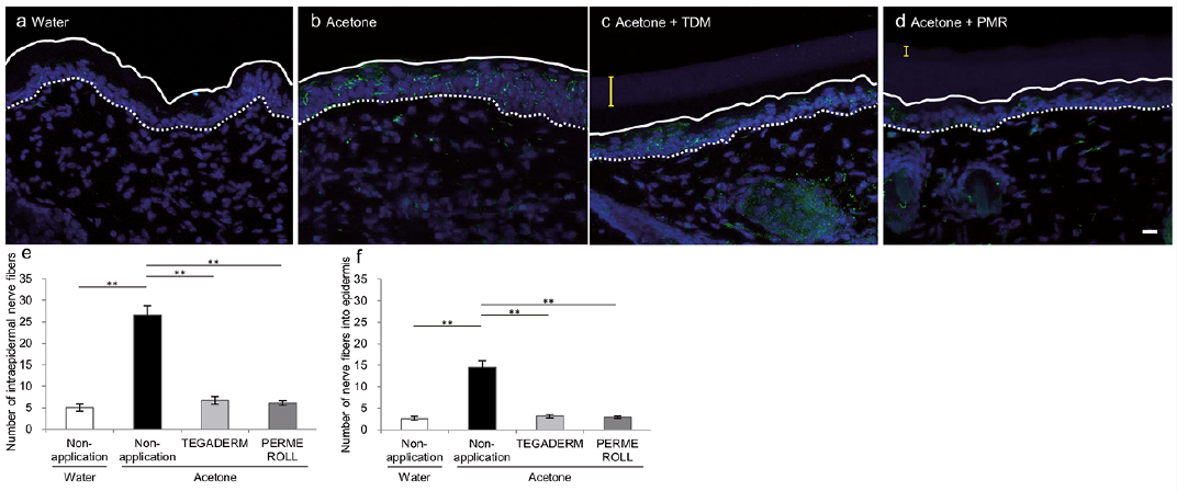
Epidermal hyper-innervation model. The numbers of PGP9.5-immunoreactive nerve fibres in and penetrating into the epidermis were significantly increased in the acetone-treated group (Fig. 1a, b, e, f). Both the number of nerve fibres in, and penetrating into, the epidermis were significantly decreased by TDM or PMR treatment (Fig. 1c–f).
Fig. 1. Effects of the immediate application of Tegaderm™(TDM) or Perme Roll™ Lite (PMR) after 5 min acetone treatment on epidermal hyper-innervation in dry skin mice. (a–d) The distribution of intraepidermal nerve fibres in the epidermis of acetone-induced dry skin mice were examined immunohistochemically with anti-protein gene product 9.5 antibody and Alexa 488-conjugated secondary antibody (green), and the nuclei were stained with DAPI (4′,6-diamidino-2-phenylindole) (blue). The white and broken lines indicate the skin surface and border between the epidermis and dermis, and yellow lines are the thickness of the film dressing. Scale bar: 12.5 µm. (e) The number of nerve fibres in the epidermis and (f) penetrating into the epidermis significantly increased in the acetone-treated group 48 h after treatment compared with that of the water-treated group (**p < 0.01), and epidermal hyper-innervation was significantly inhibited by the application of TDM and PMR. All values represent the mean and standard error of the mean (SEM) of 8 animals. Statistical analysis was performed using one-way analysis of variance (ANOVA) followed by Tukey-Kramer multiple comparisons post-test (**p < 0.01).
Alloknesis model. The AEW- or water-treated site was divided into 9 sections (Fig. S1a). Alloknesis scores on the right or left rostral section of the AEW-treated site were significantly higher than those on the same sections of the water-treated site and on the other 7 sections of the AEW-treated site (Fig. S1b). TDM or PMR with a diameter of 8 mm was applied on the alloknesis spot of the AEW-treated site on the day after 8 days of AEW treatment (Fig. S1c). TDM and PMR application significantly prevented induction of alloknesis on the right or left rostral section of the AEW-treated site Fig. S1d).
This study showed that alloknesis scores were higher on the right and left rostral sections of the AEW-treated site compared with those in other sections, which suggests that these spots are more likely to develop alloknesis in AEW-treated dry skin mice. This result is supported by a previous study showing that mechanical stimuli induces strong itch in a specific receptive field, such as a limited area between the lower lip and chin, in humans (10). We also observed epidermal hyper-innervation in both the alloknesis spot and other sections of AEW-treated dry skin mice (Iwanaga et al., unpublished observations). Although it still unclear why alloknesis “sweet spots” exist in humans and mice, characteristic differences among sensory nerves may result in induction of alloknesis.
Topical application of film dressings for 48 h prevented intraepidermal nerve growth in acetone-treated mice. Previous studies have reported that the level of nerve growth factor in the epidermis is significantly decreased by occlusion with emollients (11) or a vapour-impermeable membrane (12). This may imply that skin moisturization prevents epidermal hyper-innervation induced by barrier disruption. Taken together, application of film dressings, such as TDM and PMR, may not only induce wound healing, but also prevent epidermal hyper-innervation. In addition, film dressings may reduce itch caused by external stimuli, such as allergens (house dust mites) and clothes (wool), on skin.
This work was partly supported by a grant from the Strategic Research Foundation Grant-aided Project for Private Universities from MEXT (grant number S1311011).
The authors have no conflicts of interest to declare.


 Click to show fullsize