


1Department of Dermatology, Reims University Hospital, University of Reims Champagne-Ardenne, Avenue du general Koenig, FR-51092 Reims Cedex, 2Biopathology, Hospital Maison Blanche, Reims University Hospital, 3Department of Medical Oncology, Jean Godinot Institut, Reims, 4Department of Rheumatology, Manchester Hospital, Charleville Mézières, and 5Dermatopathology and Dermatology, Faculty of Medicine, University of Strasbourg, University Hospital of Strasbourg, Strasbourg, France. *E-mail: cvanhaecke@chu-reims.fr
Accepted Oct 3, 2018; Epub ahead of print Oct 3, 2018
Granuloma annulare (GA) is a benign inflammatory skin disease of unknown origin, classically presenting with self-limited, skin-coloured to erythematous, papules with ring-shaped distribution with no epidermal changes, often located on the dorsal surfaces of the hands and feet.
Several clinical variants of GA have been described, including generalized GA, subcutaneous GA, and perforating GA. GA shows typically palisading granulomas with a central zone of necrobiotic collagen surrounded by a palisade of histiocytes associated with mucin deposition. An interstitial histopathological pattern is also described, consisting of lymphohistiocytic infiltrate scattered between and around collagen bundles and around blood vessels in the reticular and papillar dermis. Finally, subcutaneous histopathological patterns are also reported.
With the exception of diabetes, no consistently associated systemic disorder is identified in GA, and it is not usually considered as a paraneoplastic syndrome. We report here an unusual case of diffuse interstitial generalized GA in association with breast cancer.
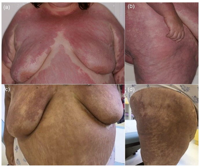
A 45-year-old woman was referred to our dermatology unit with a 4-month history of an extensive rash with large erythematous pruritic papulous plaques (Fig. 1 a, b). The lesions initially involved sun-exposed sites, and rapidly spread on the limbs, trunk and face with no eyelid involvement. The patient reported symmetrical polyarthritis of the ankles, knees and wrists, in association with high-grade fever, marked asthenia and 15 kg weight loss. No myalgia, motor deficit or synovitis was present. Physical examination revealed a voluminous left mammary tumour T2 (5 cm) and palpable axillary lymph nodes N1 (6 cm). Her medical history was marked by diabetes mellitus (DM) and dyslipidaemia. Creatine kinase and aldolase concentrations were normal, antinuclear antibodies titre was 1:400 with speckled pattern. Tests for anti-extractable nuclear antibodies, anti-double-stranded DNA, anti J0-1 antibodies, rheumatoid factor and anti-cyclic citrullinated peptide antibodies were negative. There was no specific radiological damage in the peripheral joints. Cutaneous biopsies revealed a histiocytic infiltrate, with no multinucleated giant cells, in an interstitial and diffuse pattern from the papillary to the reticular dermis, with no interface dermatitis. Immunohistochemical stains showed diffuse CD68+ and CD163+ histiocytes (Fig. 2).
Fig. 1. Clinical photographys at the time of breast cancer diagnosis, showing symmetrically large erythematous papulous plaques involving: (a) chest; (b) thigh, after 2 cycles of neoadjuvant chemotherapy for breast cancer with residual post-inflammatory pigmentation involving the same locations as the initial lesions: (c) chest and (d) thigh.
Fig. 2. Histopathological findings. (a) Haematoxylin-eosin stain (magnification ×250): lymphocytic and histiocytic (yellow arrows) infiltrate scattered between and around collagen bundles and around blood vessels with no multinucleated giant cells in an interstitial and diffuse pattern in the dermis. (b) Immunohistochemical CD163 stain (magnification ×250): interstitial and diffuse positive histiocytes.
The clinical presentation of this patient was atypical for GA, with large erythematous papulous plaques with an active border, involving a large skin surface. The lack of annular disposition was particularly puzzling; nevertheless, non-annular lesions are usual in the generalized variety of GA (1, 2). The diagnosis of multicentric reticulohistiocytosis (MRH) could also have been suspected because of concomitant polyarthritis and association with tumoural syndrome. Indeed, this rare systemic inflammatory granulomatous disease is characterized by cutaneous papulonodules and severe and rapidly progressive polyarthritis, with association with various malignancies in up to 25% of cases (3). Nevertheless, the classical MRH histopathological findings, i.e. infiltrate of histiocytic prominent multinucleated giant cells with eosinophilic “ground-glass” cytoplasm (4), were not observed in our patient’s biopsies. The association in this case of polyarthritis was also puzzling, with a strong parallel evolution of skin and joints symptoms, leading us to consider, together with the rich interstitial histiocytic infiltrate observed in the dermis, the diagnosis of interstitial granulomatous dermatitis (IGD). Nevertheless, other histopathological characteristics of IGD were not observed. The histiocytic infiltrate involved the whole height of the dermis, including the papillary dermis, while it involves only the middle and deep dermis in IGD. Moreover, the collections of histiocytes were scattered homogeneously between and around collagen bundles, and around blood vessels, while focal areas of palisading around necrotic collagen fibres are seen in IGD. Altogether, these key histopathological characteristics favoured the diagnosis of the diffuse interstitial pattern of GA (5, 6). The occurrence of arthritis has indeed been described in generalized GA (2), while we cannot exclude that it was also part of a paraneoplastic syndrome, as reported in the course of several malignant neoplasms, including breast cancer (7, 8).
Regarding the underlying conditions presented by our patient, diabetes and dyslipidaemia are diseases widely reported to be associated with GA (1, 2, 9). Importantly, a few cases of GA associated with malignant neoplasms have also been described (10–12), more specifically in generalized GA (2, 13, 14). A previous case of a GA in a 52-year-old woman, preceding by one month the diagnosis of breast cancer, resolved 1 month after mastectomy and recurred 1 month before metastasis (12). While the wide variation in the time-span between cancer and GA occurrence reported in the literature usually does not suggest a causative relationship (2, 10, 15), the delay between the onset of GA and the discovery of breast cancer in our case, was short, and the treatment of the underlying malignancy resulted in regression of the dermatological and rheumatological disorder. Spontaneous resolution of GA can be expected, but is rarely observed in generalized GA (1, 2).
We report here a case of generalized interstitial GA associated with polyarthritis, with a strong suspicion of both manifestations being paraneoplastic.
The authors have no conflicts of interest to declare.


 Click to show fullsize
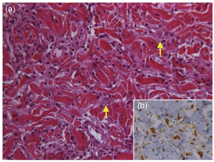 Click to show fullsize