


1Department of Dermatology and Venereology and 2Department of Pathology and Cytology, Skåne University Hospital, Scaniaplatsen 5, SE-211 17 Malmö, Sweden. E-mail: gregtheodosiou@yahoo.com, Grigorios.Theodosiou@skane.se
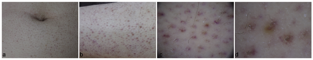
A 55-year-old man was referred to the outpatient clinic of our department with suspected vasculitis. The patient was admitted to the emergency department due to a progressive worsening of his general condition, anemia and rash. He was a homeless, unemployed, single man with a history of alcohol overconsumption. He had poor dentition without gingival bleeding. He had small, non-infiltrative perifollicular purpuric papules symmetrically distributed on his abdomen, arms and legs (Fig. 1a, b). Dermatoscopic examination highlighted the presence of coiled hairs surrounded by a purple to violaceous peripheral ring (Fig. 1c, d). Histopathology revealed the presence of extravasated erythrocytes in a perifollicular distribution, follicular hyperkeratosis with coiled, fragmented hairs in a hyperkeratotic follicular material (Fig. 2).
What is your diagnosis? See next page for answer.
Fig. 1. a, b: Perifollicular, hyperkeratotic papules. c, d: Contact non-polarized dermatoscopy: coiled hairs surrounded by a violaceous halo.
Fig. 2. a, b: Dilated follicular infundibulum with hyperkeratosis and cork screw hair. c, d: Perifollicular lymphohistiocytic infiltrate with extravasated erythrocytes without signs of vasculitis (hematoxylin-eosin stain: a: 20x, b: 100x, c, d 400x).
Acta Derm Venereol
Diagnosis: Scurvy
The clinical and histopathologic findings were consistent with the diagnosis of scurvy. The patient was treated with 1,000 mg of ascorbic acid daily. The cutaneous manifestations and coexisting anemia have resolved by the follow-up visit at 4 weeks.
Scurvy is a potentially lethal condition caused by the inadequate vitamin C intake. Ascorbic acid (vitamin C) is a water-soluble vitamin essential for the formation of normal collagen and hair growth. Scurvy is clinically characterized by spontaneous perifollicular petechiae and ecchymoses, bleeding gums, abnormal hair growth with the formation of coiled hair (´corkscrew hair´), and poor wound healing. The most common causes are malnutrition, gastrointestinal disease, poor dentition and alcoholism. Concomitant deficiencies of other factors may contribute to the appearance of the cutaneous lesions (1, 2).
Vitamin C deficiency can present at any age, although it is uncommon in the neonatal period. There is no sex or ethnic predilection. Vitamin C deficiency can also be associated with both folate and iron deficiencies (1, 3).
Individuals may complain of lethargy, fatigue, malaise, emotional lability, arthralgias, weight loss, anorexia and diarrhea. They may also experience easy bleeding, bruising, and poor wound healing. A normochromic monocytic anemia is common, resulting from blood loss, folate and iron deficiencies. Visceral or brain bleeding leads to convulsions and shock. Death may occur abruptly (1–3).
Oral disease is prominent among patients with pre-existing poor dentition. Hemorrhagic gingivitis is the most common manifestation (1, 3).
Scurvy may be misdiagnosed as leukocytoclastic vasculitis. The differential diagnosis also includes purpura, associated with various hematological conditions, collagen vascular diseases, trauma, and medications.
The major complication associated with scurvy is hemorrhage into various tissue sites, which can potentially result in a permanent functional impairment.
The diagnosis is largely clinical. Serum ascorbic levels may be measured (< 11 mmol/l), but typically reflect the recent dietary intake. Measurement of leucocyte ascorbic acid levels tends to be more accurate, but testing is not widely available (1–4).
Dermatoscopic findings include follicular hyperkeratosis, coiled hair and a perifollicular pale-orange halo (4–8).
This case illustrates that scurvy is a diagnosis, which can be easily missed. Dermatoscopy can facilitate early diagnosis and prompt therapy. Scurvy remains a disease with a potential fulminant course and even a risk of death.


 Click to show fullsize
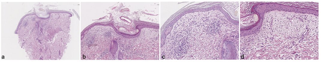 Click to show fullsize