


Division of Dermatology, Department of Medicine of Sensory and Motor Organs, Faculty of Medicine, Tottori University, 36-1, Nishi-cho, Yonago, Tottori 683-8504, Japan. E-mail: higoto@med.tottori-u.ac.jp
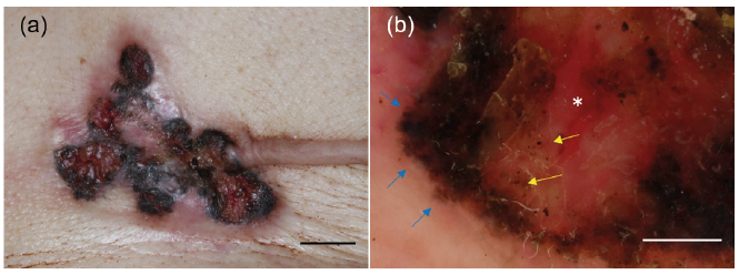
An 89-year-old woman was referred to our clinic with ulcerative pigmented nodules on the left side of her chest. She had had a nodule 3 years previously, and the number of nodules gradually increased. The size of the nodule enlarged with ulceration. She had a past medical history of breast cancer of the left breast, which had been treated surgically 4 years previously. She had not received any adjuvant therapies, including chemotherapy and radiation therapy, after the operation. Physical examination revealed multiple pigmented nodules with ulceration on the postoperative scar (Fig. 1a). These were fused to each other and the whole lesion was 4 cm in size. Dermoscopic examination showed dark-brown, leaf-like pigmented areas and ulceration with some dots/globules (Fig. 1b).
What is your diagnosis? See next page for answer.
Fig. 1. (a) Physical examination revealed pigmented nodules with ulceration on the postoperative scar (bar=1cm). (b) Dermoscopic examination revealed dark-brown leaf-like pigmented areas (blue arrows) and ulceration (*) with some dots/globules (yellow arrows) (bar=2 mm).
Acta Derm Venereol 2020; 100: adv00177.
Diagnosis: Epidermotropic recurrence of breast cancer
Skin biopsy was performed from a pigmented nodule on the chest. Histopathological examination revealed a proliferation of ductal nests composed of atypical cells in the epidermis to deep dermis. Furthermore, in some parts, epidermotropism of the tumour cells and some melanophages in the superficial dermis were observed (Fig. 2). Immunohistochemistry showed positivity of oestrogen receptor (100%) and negativity of progesterone receptor (1%). These findings were almost the same as those in tumour cells of the primary breast lesion resected previously. A diagnosis of recurrent breast cancer was made. Computed tomography revealed multiple lung metastases, and the patient agreed to best supportive care.
Pigmented recurrent breast cancer is unusual. In the case described here, the clinical manifestations were similar to those of pigmented basal cell carcinoma (BCC). BCC can occur in irradiated regions. However, the patient had not received radiation therapy after the operation. A recurrence of breast cancer was suspected, but the possibility of pigmented BCC could not be excluded based only on the dermoscopic findings.
Recurrent breast cancer occasionally showed pigmentation, and some bore clinical manifestations resembling melanoma (1–6). In some cases, dermoscopic features of melanoma including network structures and blue whitish veils were not seen. However, leaf-like areas, dots/globules and ulceration were observed, sharing dermoscopic findings of pigmented BCC. Pigmented recurrence of breast cancer often showed epidermotropism of tumour cells. In addition, such cases had colonization of melanocytes in the tumour cell nests, melanin deposition in the tumour cells, and melanophages in the superficial dermis (1, 3, 4, 6). Therefore, epidermotropism of the tumour cells seems to be a key aspect of pigmented recurrent breast cancer, giving an appearance of BCC under dermoscopy (6). It is important for dermatologists to make a diagnosis based on the results of histopathological examination.
Fig. 2. Epidermotropism of the tumour cells and some melanophages in the superficial dermis were observed (bar=100 μm).


 Click to show fullsize
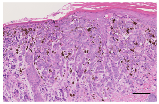 Click to show fullsize