


Departments of 1Dermatology, and 2Pathology, Pontevedra University Hospital, Centro de Especialidades Mollabao, Rúa Simón Bolívar S/N, ES-36003 Pontevedra, Spain. E-mail: carmen.couselo.rodriguez@sergas.es
Pigmented lesions are a frequent challenge for dermatologists, due the difficulty of differential diagnosis between common entities in clinical practice, such as seborrhoeic keratosis, basal cell carcinoma, melanocytic lesions and adnexal tumours. A thorough history and physical examination, always accompanied by dermoscopy, are essential.
We report here 2 cases of middle-aged patients, who were otherwise healthy, with solitary acquired pigmented lesions, who were referred to our dermatology department in order to rule out malignant melanoma.
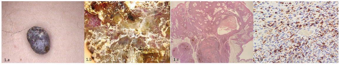
Case 1. A 55-year-old man presented with an excrescent, dark-brown lesion, 13 mm in diameter located on his right shoulder. Dermoscopy revealed irregular polymorphous vascularity, blue-grey ovoid nests, and keratinization. The patient reported that the lesion had grown in the last years. The lesion was excised and sent to the pathology department (Fig. 1).
Case 2. A 65-year-old woman with a firm, slow-growing, black nodule, 15 mm largest diameter, located on the inner side of her left thigh was referred to our unit. Dermoscopy revealed blue-grey pigmentation, globule-like structures, comedo-like structures, irregular vascularity and keratinization. The lesion was removed and sent for histology (Fig. 2).
What is your diagnosis? See next page for answer.
Fig. 1. Case 1. (a) Pediculated, dark-brown lesion located on the patient’s right shoulder. (b) Dermoscopy reveals irregular polymorphous vessels, blue-grey ovoid nest, and keratinization. (c) Well-defined dermal tumour connected to epidermis, heavily pigmented. Haematoxylin and eosin (H&E), original magnification 40×. (d) Cytoplasmic positive immunostaining with HMB45, original magnification 200×.
Fig. 2. Case 2. (a) Slow-growing black nodule, located on inner face of the patient´s left thigh. (b) Dermoscopy shows blue-grey pigmentation, globule-like structures, comedo-like structures, irregular vascularity and keratinization. (c) Sharply demarcated celular proliferation with cytoplasmic melanin granules. H-E, 100x. (d) Positive staining with HMB45 in dendritic cells. HMB45, 400x.
Acta Derm Venereol 2020; 100: adv00195.
Diagnosis: Pigmented eccrine poroma
Histopathological examination of case 1 revealed a well-defined cellular proliferation with features of poroid differentiation (Fig 1c); melanin pigment within the cytoplasm of the tumoural cells was observed, with some cells showing immunostaining with HMB45 (Fig. 1d). In case 2 a sharply demarcated dermal tumour formed by monomorphic cuboidal cells with oval nuclei and occasional cytoplasmic melanin granules was observed (Fig. 2c). Dendritic melanocytes without atypia were scattered evenly within the tumour, showing immunostaining with both HMB45 (Fig. 2d) and S100. Immunostaining with AE1/AE3 was also performed, demonstrating staining of epithelial neoplastic cells. Atypical mitoses, cytologic atypia or other features suggestive of malignancy were not seen. Based on these findings, a diagnosis of pigmented eccrine poroma was established in both patients.
Eccrine poroma is a benign adnexal neoplasm described by Pinkus in 1956. Conventional eccrine poroma is commonly presented as a flesh-coloured to reddish nodule, papule or plaque, at the acral sites, which are the sites with higher concentration of eccrine sweat glands. It is more frequent between the 4th and 6th decades of life, without sex predilection (1).
The pigmented variant of the eccrine poroma is rare and has been poorly reported in the literature (2). This variant seems to be more frequent in non-white people and on non-acral sites (3). At clinical examination the lesions appear as well-circumscribed papules, plaques and nodules, brown to black in colour. Clinical differential diagnosis includes pigmented basal cell carcinoma, nodular melanoma, pigmented porocarcinoma and seborrhoeic keratosis (4).
In addition to being clinically similar to a range of other benign and malignant tumours, pigmented poroma is also considered a great dermatoscopic imitator. Hence, in dermoscopy we can find blue-grey ovoid nest and arborizing vessels mimicking a basal cell carcinoma (5), and we can observe comedo-like openings and hairpin vessels simulating a seborrhoeic keratosis, or blue-whitish veil, setting differential diagnosis with malignant melanoma (6). Minagawa & Koga (6) observed that the most frequent dermatoscopic structures in pigmented poromas were vascular structures (hairpin vessels, polymorphic and arborizing vessels) followed by globule-like structures. To date, no pigment network has been described as a dermatoscopic finding.
Therefore, since pigmented poroma can simulate multiple tumours the diagnosis of certainty is histological, showing solid nodules of uniform small round cells and admixed dendritic melanocytes (1) that are responsible for the dark clinical appearance, together with melanophages within adjacent stroma (3).
Recurrences or even malignant progression into eccrine porocarcinoma (7, 8) have been reported, therefore surgical excision is the treatment of choice for pigmented poroma.


 Click to show fullsize
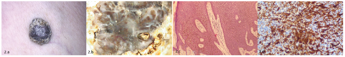 Click to show fullsize