


Ülker Gül, Arzu Kılıç, Müzeyyen Gönül, Seray Külcü Çakmak and Ceren Erİnçkan
2nd Dermatology Clinic, Ankara Numune Education and Research Hospital, Ankara, Turkey
Cutaneous metastases may be either the initial manifestation of an internal malignancy or represent recurrent neoplastic disease. The aim of this study was to investigate the incidence and characteristics of cutaneous metastases in cases of internal malignancy. A total of 1287 patients with internal malignancy were included in the study. Dermatological examinations were performed on all of the patients. Skin biopsies were obtained from the suspected lesions. The type of malignancy, the time of diagnosis of the malignancy, the presence of cutaneous metastasis, and the localization sites of the cutaneous malignancy were noted. Metastases of any kind were seen in 27.4% of cases. Cutaneous metastases were seen in 1.2% of cases and were most frequently localized on the anterior chest as nodules. Cutaneous metastasis was the first sign of internal malignancy in one case. It is concluded that cutaneous metastases occur rarely and the presentation of internal malignancy with skin involvement is uncommon. Key words: cutaneous; metastasis; malignancy.
(Accepted September 26, 2006.)
Acta Derm Venereol 2007; 87: 160–162.
Arzu Kılıç, Onur Sok. No. 47/11, TR-06570 Anittepe-Ankara, Turkey. E-mail: kilicarzu2004@yahoo.com
Cutaneous metastases of internal malignancies are not observed very often. Although cutaneous metastasis may represent the first sign of malignancy, it can be observed at any stage of malignancy (1). Early detection of cutaneous metastasis is very important, not only for diagnosis, but also for treatment of the primary tumour (1–3). The aim of this study was to examine the incidence and characteristics of cutaneous metastases of internal malignancies.
MATERIALS AND METHODS
Patients diagnosed with an internal malignancy between 2001 and 2004 in the Oncology Education and Research Hospital were consecutively included in our study. Physical and dermatological examinations were performed on all of the patients. The patients were fully undressed during the examination. Skin biopsies and histopathological examinations were performed from suspected lesions. The patients were followed up until their biopsy results were obtained within a couple of weeks. The type of malignancy, the time of diagnosis of the malignancy, the period between the appearance of the primary malignancies and cutaneous metastases were obtained from the records. The presence of cutaneous metastasis and the localization sites of the cutaneous metastases were noted.
RESULTS
A total of 1287 patients with internal malignancies were evaluated: 555 (43.1%) were female and 732 (56.9%) were male (age range 3–82 years (mean 46.5)). Two hundred and forty-five (19%) had malignancies of the gastrointestinal system, 212 (16.5%) of the lung, 198 (15.4%) of the breast, 190 (14.8%) of the genitourinary system, 173 (13.4%) of the head-neck, 152 (11.8%) of the haematological system, 80 (6.2%) of bone, 23 (1.8%) of the brain and 14 (1.1%) had other malignancies. Metastases (both internal and cutaneous) were seen in 352 (27.4%) cases. These were distributed as follows: 106 (30.1%) to lymph nodes; 74 (21.0%) to lung; 66 (18.8%) to liver; 61 (17.3%) to bone; 30 (8.5%) to brain; 15 (4.3%) to skin; 6 (1.7%) to bone-marrow; 4 (1.1%) to oesophagus; 29 (8.2%) to other organs.
Cutaneous metastases were seen in 15 (1.16%) of all cases with malignancies. The internal malignancies underlying cutaneous metastases are listed in Table I.
Table I. Internal malignancies with cutaneous metastases in order of prevalence
| Malignancy | Total frequency (n) | Frequency of internal metastases (n) | Frequency of cutaneous metastases n (% out of internal metastases/% out of all cases) |
| Haematological | 152 | 24 | 4 (16.6/2.6) |
| Breast | 198 | 68 | 5 (7.4/2.4) |
| Lung | 212 | 59 | 4 (6.8/1.9) |
| Gastrointestinal system | 245 | 69 | 2 (2.9/0.8) |
Cutaneous metastases were most frequently (2.6%) seen in cases with haematological malignancies. Six of 15 cases (40%) with cutaneous metastases had also internal metastases. When studied with respect to gender, cutaneous metastases were most frequently observed in breast cancer in females and lung cancer in males.
Nodules were the most frequent presentation (Fig. 1). Cutaneous metastasis was the first sign of internal malignancy in one patient and internal malignancy and cutaneous metastasis were diagnosed simultaneously in another patient with breast cancer. In other cases, cutaneous metastases were diagnosed after the recognition of internal malignancy. Six cases metastasized to the anterior chest wall (4 breast, 1 lung, 1 non-Hodgkin’s lymphoma), 4 to the lateral trunk (1 breast, 3 lung), 2 to the scalp (1 colon, 1 chronic lymphocytic leukaemia (CLL)), 2 to the shoulder (1 oesophagus, 1 CLL), 3 to the abdomen (2 acute myelocytic leukaemia (AML), 1 CLL), and 3 to the upper extremities (2 AML, 1 CLL) (Table II). The period between the diagnosis of internal malignancies and cutaneous metastases varied between 2 and 84 months, except in the 2 cases where the skin findings heralded a diagnosis of internal malignancy.
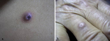
Fig. 1. Nodular lesion of (a) lung adenocarcinoma on the trunk and (b) chronic lymphocytic leukaemia on the finger.
Table II. Localization of cutaneous metastases in various internal malignancies
| Malignancy with cutaneous metastases | No. of cutaneous metastases | ||||||
| Anterior chest wall | Lateral trunk | Scalp | Shoulder | Abdomen | Upper extremities | Total | |
| Haematological | 1 | 0 | 1 | 1 | 3 | 3 | 9a |
| Breast | 4 | 1 | 0 | 0 | 0 | 0 | 5 |
| Lung | 1 | 3 | 0 | 0 | 0 | 0 | 4 |
| Gastrointestinal system | 0 | 0 | 1 | 1 | 0 | 0 | 2 |
aNine metastases in 4 patients
DISCUSSION
The diagnosis of cutaneous metastasis and the underlying tumour has significance for both the patient and the clinician. The discovery of cutaneous metastasis in a patient with a known primary tumour may be the first sign of recurrence (1–3). Moreover, cutaneous metastatic growths may change the therapy regimen (4).
In the literature, the incidence of cutaneous metastatic disease was reported to be between 0.7% and 9% in cases with cancer (1). We found a total of 15 cases of skin involvement among 1287 cases of carcinoma (1.16%). In our study the cutaneous metastases accounted for 1,2% of all cases, which is lower than the average 2.3% reported in previous retrospective and autopsy studies (1).
Although cutaneous metastases are generally detected during the advanced stages of malignancies, they can occasionally be the first sign of a visceral cancer (2). Brownstein et al. (4) reported that lung, kidney and ovary cancers are the most common type of cancers with skin involvement as the presenting sign. They indicated that 3% of cases of carcinoma of breast presented with cutaneous metastases. Lookingbill et al. (1) reported that 1.3% of the cases had skin involvement at presentation of cancer. In our study cutaneous metastatic disease preceded the diagnosis of breast cancer in one case, and besides cutaneous metastatic disease and breast cancer, were diagnosed simultaneously in another case.
The cancer types that most commonly metastasize to skin are lung and colon cancers in males, and breast, colon and ovary cancers in females (1, 4, 5). In our study, cutaneous metastatic disease was detected most commonly in lung carcinoma in males and breast carcinoma in females.
Cutaneous metastases may be the result of direct invasion, local metastasis or distant metastasis from the primary site (1). In our study, 4 of the cases had direct extension of the tumour to the skin, 5 had local metastases and 6 had distant metastases. Four of the direct extensions were breast cancers. Four of the distant metastases were haematological malignancies, one was colon cancer, and the other was oesophagus cancer. Four of the local metastases were lung cancers, and the other one was breast cancer. Five of the distant metastases had nodular presentation, one had cicatriciel type.
Cutaneous metastases show predilection for certain regions (1, 2, 4). Breast and lung cancers frequently metastasize to the chest wall, whereas cancers of the bowel, ovary, and bladder most often metastasize to the abdomen. The localization sites of the cutaneous metastasis in our study were to the anterior part of the chest in 6 cases, 4 of which had breast cancer. There were 4 cases that metastasized to the lateral trunk, 3 of which had lung cancer and one had breast cancer. There were also cutaneous metastases in 2 patients to the scalp (colon adenocarcinoma, CLL), and to the shoulders, abdomen and upper extremities. The scalp is a relatively rare site for the localization of cutaneous metastases (4) that may be clinically atypical (6). Cutaneous metastases can occur solitarily or multiply (2). The lesions may present as dermal papules, subcutaneous nodules, inflammatory patches, or as fixed, indurated lesions. The most common presentation is as multiple nodular lesions (3). We have observed nodular metastases in 13 of our patients (4 solitary, 9 multiple). This finding is consistent with the literature. One of the patients had cicatricial alopecia, previously described in a case report (7), and one had peau d’orange (breast carcinoma). All of the haematological malignancies had nodular metastases; one of was solitary (non-Hodgkin’s lymphoma), the others were multiple nodules (2 AML, 1 CLL).
In conclusion, this prospective study shows that cutaneous metastases occur infrequently and that internal malignancy rarely presents with skin involvement. However, despite a low risk of cutaneous metastasis, skin nodules of an unknown cause should be biopsied frequently to detect these uncommon, but sometimes very early, manifestations of internal malignancy.
REFERENCES
1. Lookingbill DP, Spangler N, Sexton FM. Skin involvement as the presenting sign of internal carcinoma. A retrospective study of 7316 cancer patients. J Am Acad Dermatol 1990; 22: 19–26.
2. Schwartz RA. Cutaneous metastatic disease. J Am Acad Dermatol 1995; 33: 161–182.
3. Lookingbill DP, Spangler N, Helm KF. Cutaneous metastases in patients with metastatic carcinoma: A retrospective study of 4020 patients. J Am Acad Dermatol 1993; 29: 228–236.
4. Brownstein MH, Helwing EB. Patterns of cutaneous metastasis. Arch Dermatol 1972; 105: 862–868.
5. Cohen PR. Skin clues to primary and metastatic malignancy. Am Fam Phycian 1995; 51: 1199–1204.
6. Scheinfeld N. Review of Scalp Alopecia due to a clinically unapparent or minimally apparent neoplasm (SACUMAN). Acta Derm Venereol 2006; 86: 387–92.
7. Gül Ü, Kilic A, Akbas A, Aslan E, Demiriz M. Alopecia neoplastica due to metastatic colon adenocarcinoma. Acta Derm Venereol, in press.

