


Yumi Kambayashi, Taku Fujimura, Masaya Ishibashi, Takahiro Haga and Setsuya Aiba
Department of Dermatology, Tohoku University Graduate School of Medicine, Sendai, Japan. E-mail: tfujimura1@mac.com
Accepted Feb 4, 2013; Epub ahead of print Apr 19, 2013
Eosinophilic cellulitis (EC) is described as erythematous plaques with perivascular and interstitial infiltration of eosinophils. EC is associated with various skin disorders, and occurs in adverse drug reactions to many medications (1). A previous report described a patient with EC who was found to have increased interleukin (IL)-5-producing circulating T cells (2), suggesting the co-existence of a signal transducers and activator of transcription (STAT) 6-mediated Th2 response. We describe here a case of EC induced by administration of interferon (IFN)-β.
Case report
A 68-year-old man presented with a 10-year history of a black nodule on his forearm. Physical examination revealed a black, erosive, dome-shaped nodule, 20 mm in diameter, on the extensor surface of his right forearm (Fig. 1A). Excisional biopsy showed a dense infiltration of spindle-shaped atypical cells with pigmentation from the papillary dermis to the deep dermis. Immunohistochemical staining revealed that these tumour cells were positive for HMB45, S-100 and MelanA, and negative for AE1/AE3. From these results, the patient was diagnosed with malignant melanoma. The tumour was resected with a 2.5-cm surgical margin. After surgical treatment, DAV-Feron therapy (dacarbazine, nimustine, vincristine intravenously and interferon-beta injected around the scar intracutaneously) was administered once a month, for 6 months. However, during the third cycle of combination therapy, the patient reported pruritic erythematous swelling around the IFN-β injection site on the right forearm (Fig. 1B). Biopsy specimens revealed a dense infiltration of eosinophils in the superficial and deep perivascular dermis, and in the subcutaneous layer (Fig. 2A, B). Serum myeloperoxidase anti-neutrophil cytoplasmic antibodies (ANCA) were negative, and there were no clinical signs of asthma. From the above findings, Churg Strauss syndrome and eosinophilic vasculitis were excluded. Clobetasol propionate ointment 0.05% were administered twice a day, and the pruritic erythema disappeared a few days after treatment. To further investigate the immunological profiles of the infiltrating cells, immunohistochemical staining for pSTAT1 and pSTAT6 were carried out, and revealed that the infiltrating lymphocytes contained both pSTAT1+ and pSTAT 6+ cells (Fig. 2C, D). From the above data the patient was diagnosed with eosinophilic cellulitis caused by the subcutaneous administration of IFN-β. To prevent progression of the tumour, we continued to administer IFN-β. As expected, the administration of IFN-β caused pruritic erythematous swelling at the injection site every time, although the eruption was easy to control.
Fig. 1. (A) A black, erosive, dome-shaped nodule 20 mm in diameter on the right forearm. (B) Pruritic erythematous swelling around the injection site for interferon-β on the right forearm.
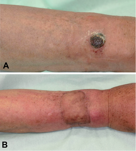
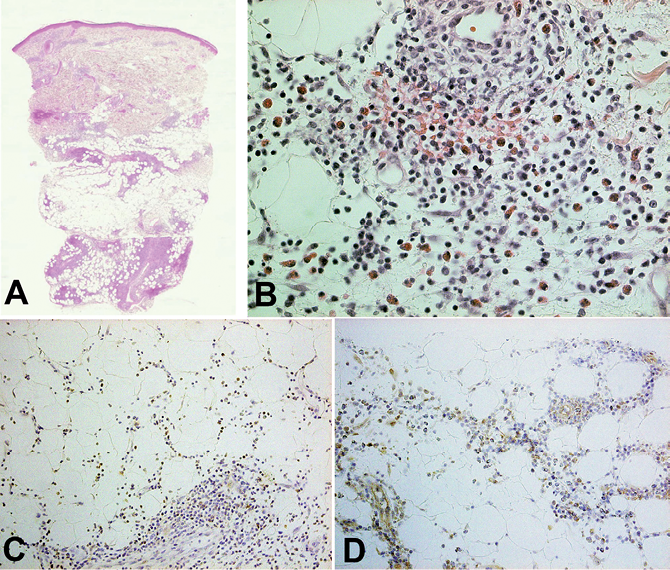
Fig. 2. (A, B) Dense infiltration of eosinophils in the superficial and deep perivascular dermis, and subcutaneous layer (original magnification (A) × 5, (B) × 400). (C, D) Paraffin-embedded tissue sample was deparaffinized and stained using (C) anti-pSTAT1 Ab, and (D) anti-pSTAT6 Ab. The sections were developed with 3,3-diaminobenzidine tetrahydrochloride (original magnification (C, D) × 200).
Discussion
IFN-β is thought to play an important role in tumour suppression in malignant melanoma (3, 4). We reported previously that peritumoural administration of IFN-β induced a significant number of CD4+ cells, CD8+ cells and TIA-1+ cells in metastatic melanoma (4). These data suggest that local administration of IFN-β induces both helper T cells and cytotoxic T cells, although precise profiles of these induced lymphocytes were not obtained. More recently, IFN-β was reported to enhance the expression of IL-12p35 and IL-12p40 mRNA on human monocyte-derived dendritic cells (MoDC) and induce the production of IFN-γ from T cells (5). In aggregate, IFN-β induces a Th1-mediated immune response at the injection site.
STATs are latent transcription factors that reside in the cytoplasm until activated (6). STAT1 is activated by Th1 cytokines such as IFN-α/β, IFN-γ and IL-27 (7), whereas STAT6 is activated by Il-4 (8). STAT6 participates in Th2 differentiation by enhancing the expression of the master regulator of Th2 differentiation, GATA3 (8). STAT6 and GATA3, together with IL-2-mediated STAT5 activation, induce the secretion of copious amounts of IL-4, IL-5 and IL-13 by activated Th2 cells (9).
EC is associated with various skin disorders, and occurs as a result of adverse drug reactions to many medications (1). In general, injection site reactions have been described as Th1-mediated reactions. Winfield et al. (1) reported a rare case of EC-like reaction caused by subcutaneous etanercept injection, which is thought to induce Th2 immune response. A previous report also described that a patient with EC showed IL-5-producing circulating T cells (2), which suggested the co-existence of a STAT6-mediated Th2 response.
We describe here a case of EC induced by the administration of IFN-β. In addition, immunohistochemical staining for pSTATs revealed dense infiltration of pSTAT1+ cells and pSTAT6+ cells in the deep dermis. To confirm the possible mechanisms of immunological profiles of infiltrating cells, we employed immunohistochemical staining for pSTAT1 and pSTAT6 for 3 cases of ordinary, idiopathic EC (data not shown). For staining pSTAT1, as we expected, few pSTAT1+ cells were observed in these 3 cases. In contrast to pSTAT1, pSTAT6+ cells were densely infiltrated around the eosinophils in the deep dermis. These data suggest that, in the present case, EC was induced by pSTAT6. Interestingly, a previous report also suggested that IFN-β inhibits Th1 polarization and promotes IL-10-producing T cells through matured DC-mediated pathways (10). Thus, in our case, repeated local injection of IFN-β may have generated matured DC at the injection site, leading to the development of EC. To our knowledge, there are no previously published reports in English of EC induced by IFN-β, and the case presented here suggests that local administration of type 1 interferon may induce EC.
References

