


Hikari Otake, Atsushi Otsuka*, Yumi Nonomura, Natsuko Iga and Kenji Kabashima
Department of Dermatology, Kyoto University Graduate School of Medicine, 54 Shogoin-Kawara, Sakyo-ku, Kyoto 606-8507, Japan. *E-mail: otsukamn@kuhp.kyoto-u.ac.jp
Accepted Sep 30, 2015; Epub ahead of print Oct 6, 2015
Papuloerythroderma is a pruritic eruption of flat-topped papules in a background of erythroderma that characteristically spares skin folds; so-called “deck-chair sign” (1). Histopathology reveals a non-specific spongiotic dermatitis-like pattern, such as slight epidermal hyperplasia with spongiosis and a primarily perivascular dermal infiltrate with lymphocytes, histiocytes and eosinophils. Peripheral eosinophilia and elevated serum immunoglobulin E (IgE) are detected in most cases of papuloerythroderma.
Recent studies have shown that basophils play some roles in the pathogenesis of cutaneous allergic diseases, where eosinophils are present, by functioning as initiator cells. CD63, CD69, and CD203c are basophil activation markers that are known to be upregulated by cross-linking of the FcɛRIα receptor. They may serve as useful activation markers in several diseases, including chronic urticaria and prurigo (2). A previous study revealed that basophil-derived interleukin (IL)-4 promotes antigen-specific T helper (Th)2 cell differentiation (3). However, it remains unknown whether basophils mediate the pathological mechanisms in papuloerythroderma. We report here 2 cases of papuloerythroderma that exhibited infiltration of basophils with upregulation of activation markers.
CASE REPORTS
Case 1. A 73-year-old Japanese man with no relevant personal history presented to our department with a 1-year history of pruritic eruptions over his trunk and all of his limbs. Physical examination revealed erythematous plaques that avoided the skin folds, and crusty indurated lesions on the abdomen (Fig. 1A). His medication was stopped in order to exclude drug eruption. Laboratory data revealed: haemoglobin 12.3 g/dl (normal range 10.2–14.6 g/dl); white blood cell count 6,500/mm3 (normal range 2,800–8,300/mm3) (61.6% neutrophils [46–62%], 17.6% lymphocytes [30–40%], 6.0% monocytes [ 4–7%], 15.2% eosinophils [3–5%] and 0.2% basophils [0–1%]); platelet count 214,000/mm3 (normal range 123,000–343,000/mm3); and immunoglobulin E (IgE) 1,900 IU/ml (normal range 0–250 IU/ml). No atypical cells were detected in peripheral blood. Computed tomography (CT) did not reveal any lymphadenopathy, splenomegaly, or visceral tumours. Oesophagogastroduodenoscopy and colonoscopy did not reveal any malignancy. A skin biopsy from an eruption on the patient’s abdomen revealed non-specific inflammatory changes with a perivascular infiltration of lymphohistiocytic cells intermingled with eosinophils in the upper dermis (Fig. 1B). Based on clinical and pathological findings, his eruptions were diagnosed as papuloerythroderma. Treatments with narrowband ultraviolet B (UVB) and topical corticosteroids gave partial relief for the pruritus and eruptions.
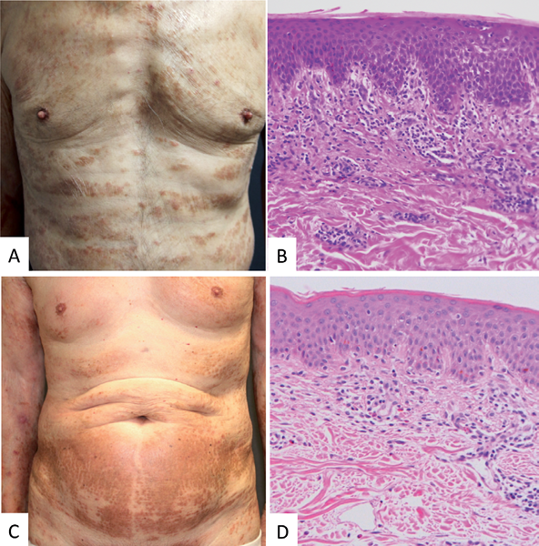
Fig. 1. (A) Papuloerythroderma with typical avoidance of abdominal skin folds. (B) Epidermal acanthotic hyperplasia with mild spongiosis. In the dermis, perivascular infiltration of lymphohistiocytic cells intermingled with eosinophils. (C) Skin lesion formed by the coalescence of flat-topped, red-to-brown papules showing the deck-chair sign. (D) Spongiosis and mild acanthosis in the epidermis together with perivascular infiltration of lymphohistiocytic cells intermingled with eosinophils. (Haematoxylin-eosin stain; original magnification × 200).
Case 2. A 74-year-old Japanese man presented to our department with a 2-month history of pruritic eruptions over his trunk and all of his limbs. He had a history of chronic hepatitis C virus infection and had had an operation for gastric cancer one year previously. He received no medication at that time. Physical examination revealed erythematous plaques that avoided the skin folds, and crusty indurated lesions on the abdomen (Fig. 2A). Laboratory data showed peripheral eosinophilia: white blood cell count 6,600/mm3 (55.4% neutrophils, 20.7% lymphocytes, 6.5% monocytes, 16.3% eosinophils and 1.1% basophils). No atypical cells were detected in the peripheral blood. CT did not show any lymphadenopathy, splenomegaly, or visceral tumours. There was no recurrence of gastric cancer. A skin biopsy from an eruption on the patient’s abdomen revealed a perivascular infiltration of lymphohistiocytic cells and eosinophils in the upper dermis (Fig. 2B). Based on these findings, his eruptions were diagnosed as papuloerythroderma. Treatments with narrowband UVB, topical corticosteroids and topical tacrolimus improved the skin lesions moderately.
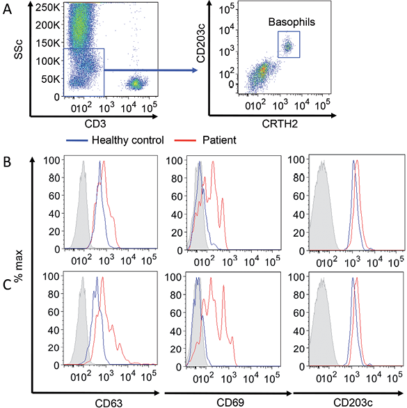
Fig. 2. (A) Gating strategy for basophils. (B) Histogram of each maker of basophil in patient 1, and patient 2 in comparison with (C) healthy donor (n = 4).
The expression levels of CD63, CD69, and CD203c on basophils in peripheral blood were evaluated before treatment by flow cytometry. Basophils were detected as CD3– CTRH+ cells (Fig. 2A). The expression levels of CD63, CD69, and CD203c on basophils in case 1 (Fig. 2B) and case 2 (Fig. 2C) were upregulated compared with those in healthy donors (n = 4). In addition, infiltration of basophils into the lesion skin was found in both cases (Fig. 3).
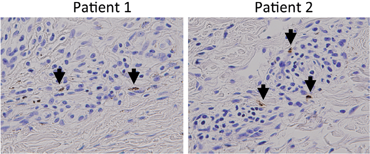
Fig. 3. Basophils were detected using anti-human basophils antibody, 2D7 (purchased from BioLegend, San Diego, CA, USA), in patients 1 and 2 (arrows). Immunostaining × 400.
DISCUSSION
The aetiology of papuloerythroderma remains largely unknown. It has been suggested to be a reaction to an unidentified cutaneous antigen, or there may be an association with neoplasia or drugs (4). Although peripheral eosinophilia and elevated serum IgE were detected in most cases of papuloerythroderma, there has been no analysis of the role of Th2 immune cells in the pathogenesis of papuloerythroderma thus far. To the best of our knowledge, this is the first demonstration that peripheral basophils are activated in patients with papuloerythroderma. Basophils release multiple mediators (e.g. leukotrienes, proteases, and cytokines, such as IL-4) that might be linked directly to Th2 skewing in the patient’s papuloerythroderma. Although the number of cases is limited, our results may shed light on the possibility that basophils play a role in the pathogenesis of papuloerythroderma.
The authors declare no conflicts of interest.
REFERENCES

