


Rudolf Happle1 and Aïcha Salhi2
1Department of Dermatology, Freiburg University Medical Center, DE-79104 Freiburg, Germany, and 2Service de Dermatologie, Hôpital central de l’Armée, Algiers, Algeria. E-mail: rudolf.happle@uniklinik-freiburg.de
Accepted Apr 14, 2016; Epub ahead of print Apr 19, 2016
Sparing of the nipple-areola complex is a typical feature of large congenital melanocytic naevi (MNs) involving the breast. In 1995, this phenomenon was comprehensively depicted in German by Konrad Bork (1) and in 2015 it was reported in English by Baykal et al. (2) who were unaware of Bork’s description. Thereupon, we asked ourselves whether an analogous sparing phenomenon may be noted in other regions of the body (3). We describe here 2 infants whose giant MN involved the abdomen but spared the umbilicus. A literature search yielded several similar cases, suggesting that this is not a purely coincidental feature of such naevi.
CASE REPORTS
Case 1. A 21-month-old girl presented to the author (AS) with a giant MN covering her back, left thigh and abdomen, with the exception of the right hypochondriac region, from which a narrow band of uninvolved skin reached the right groin. The umbilicus was partially spared, although it was completely surrounded by the naevus (Fig. 1). Many small melanocytic naevi were disseminated over her entire integument, and a large lesion, 5 cm in diameter, involved her left cheek. We advised surgical removal of the large facial lesion and regular follow-up appointments because of the risk of malignant degeneration.

Case 2. A 10-day-old boy with a giant MN was examined by the author (AS). The naevus covered most of the patient’s back, including the buttocks, the upper third of his left thigh, and most of his abdomen. Only the right hypochondriac region and a contiguous band reaching the groin remained uninvolved. The umbilicus was completely spared, whereas the structures surrounding the umbilicus were involved by the naevus (Fig. 2). Moreover, multiple disseminated small naevi involving his entire body, including the palms and soles, were noted.
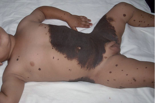
Fig. 2. Case 2: a 2-year-old boy with giant melanocytic naevus involving the abdomen but sparing the umbilicus.
DISCUSSION
To date, sparing of the umbilicus, as noted in the present cases of giant MN, has been regarded as a fortuitous finding, and thus has not been mentioned in previous reports. A literature search, however, revealed 7 reports containing photographs that documented a similar phenomenon (Table I). Umbilical sparing was complete in 4 cases and incomplete in 3 children. Remarkably, the phenomenon was documented in a 7-year-old boy, who showed, in addition, bilateral sparing of the nipple-areola complex (6).
Table I. Additional cases of giant melanocytic naevi showing umbilical sparing, as documented inadvertently on photographs published during 2002 to 2015
|
Case No. |
Authors |
Year |
Sex |
Age |
Extent of sparing |
|
1 |
Silfen et al. (4) |
2002 |
F |
8 months |
Partial |
|
2 |
Ostertag et al. (5) |
2006 |
F |
7 weeks |
Partial |
|
3 |
Kobayashi et al. (6) |
2006 |
M |
7 years |
Complete |
|
4 |
Aoyagi et al. (7) |
2007 |
F |
2 months |
Partial |
|
5 |
Konz (8) |
2007 |
M |
3 weeks |
Complete |
|
6 |
Coyette et al. (9) |
2014 |
F |
24 days |
Complete |
|
7 |
Sawicka et al. (10) |
2015 |
M |
4 months |
Complete |
How can this sparing phenomenon be explained? Similar to the nipple-areola complex, the tissue of the umbilicus appears to be a less suitable habitat for the cells of large MNs immigrating into the skin during embryogenesis. Remarkably, small umbilical MNs have site-specific histopathological characteristics, such as prominent lamellar fibrosis (11). It should be emphasized, however, that large MN may also involve the umbilicus.
Current data suggests that sparing of the umbilicus may represent a characteristic feature of giant MNs. Further clinical reports may show whether our assumption holds true. From a theoretical point of view, this sparing phenomenon may be important for the understanding of the aetiology and pathogenesis of large MN. As a possible practical implication, one may speculate that both umbilicus and nipple-areola complex, if involved by a large MN, are less prone to develop malignant melanoma. Interestingly, there are reports on primary umbilical melanoma (12), but we were not able, so far, to find a case of umbilical melanoma originating from a large MN.
REFERENCES

