


1CIR-Dental School, Department of Surgical Sciences, University of Turin, Via Nizza 230, 10126 Turin, 2Torino Cord Blood Bank – Immunohematology and Transfusional Medical Service, A.O.U. Città della Salute e della Scienza, Turin, Italy, and 3World Health Organization Collaborating Centre for Oral Health-General Health, UK. *E-mail: paologiacomo.arduino@unito.it
Accepted Aug 16, 2016; Epub ahead of print Aug 18, 2016
Inherited epidermolysis bullosa (EB) is a rare group of genetically heterogeneous diseases, characterized by deficiencies in the adhesion of the connective tissue to the epithelium. Junctional EB and dystrophic EB, are the most severe types. Common clinical manifestations are mechanical fragility of the skin and mucosae, with blister formation and abnormal wound healing, the severity of the lesions depending on the distinct type (1–3). The oral cavity is frequently involved, but, although bullae and erosions are common, there is only one published study of therapy, reporting that sucralfate suspension can reduce the development and duration of blisters and ulcers, reduce the associated pain, and improve indices of gingival inflammation (4).
Autologous and allogeneic platelet (PLT) preparations, in particular a blood component termed “platelet gel”, traditionally obtained from adult blood platelets, are rich in regenerative growth factors, which are valuable for the treatment of chronic wounds (5). Recent findings on multiple biological properties of human umbilical cord blood (CB), and its high level of viral safety, prompted one group to produce PLT gel from CB (cord blood platelet gel; CBPG) (6), and another to detail its effectiveness for the treatment of EB skin lesions (7).
Low-level laser therapy (LLLT) has potential biostimulating effects, improving wound healing, and is a possible treatment for autoimmune oral erosive lesions, with a notable analgesic effect (8).
We therefore performed a pilot evaluation of the efficacy and safety of CBPG with LLLT for the treatment of inherited EB oral mucosal lesions, over a 3-day treatment period (one application each day).
Patients with dystrophic EB and symptomatic oral lesions were prospectively selected between June and October 2015. The ethics review board of the “Azienda Ospedaliera Città della Salute e della Scienza of Turin”, Turin, Italy, approved the study (protocol number 0089210_CS/585/09-2015). Participants with long-standing ulcerations were selected.
CB units not fulfilling the criteria for banking for transplant purposes were processed within 48 h of collection to obtain CBPG according to the “Italian Cord Blood Platelet Gel project” protocol (9) (for complete details see Appendix S1).
Morphological changes were measured with a 15-mm periodontal probe (PCPUNC15: Hu-Friedy®, Chicago, IL, USA).
The symptom score (for reporting pain) was recorded using a visual analogue scale (VAS); patients were requested to mark the scale at each visit, before and after the laser session.
The primary outcome measures were lesions’ size and pain; secondary outcome measure was adverse effects. All data were collected during a 6-month follow-up period (at 1 (T4), 4 (T5), 12 (T6) and 24 (T7) weeks after the end of the therapy).
Wilcoxon’s signed rank was used to calculate the differences of the outcome data. p-values 0.05 were considered statistically significant. SPSS for Windows (version 11, SPSS Inc., Chicago, IL, USA) statistical software was utilized.
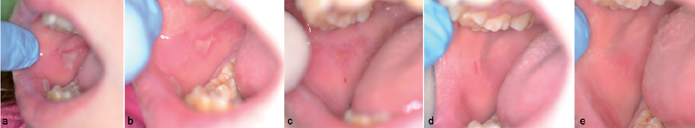
Seven patients (4 males) with dystrophic EB were included. The mean age at baseline was 19.8 years (range 8–34). Nineteen oral lesions were treated; the buccal mucosa was the most common site (37%) (Fig. 1), followed by the lips (27%), tongue (21%), floor of the mouth (11%) and gingiva (4%).
Fig. 1. Oral lesions in dystrophic epidermolysis bullosa. Case 3: A right buccal mucosal lesion at (a) T1_day1, (b) T2_day2, (c) T3_day3, (d) T4_day7, and (e) T7_24 weeks after end of therapy.
A statistically significant difference was observed for reported pain and clinical size of lesions from the first day of treatment provided (Table I). Statistical analysis was performed only until T4. At T5 and T6 no patients presented lesions in the treated area. During the follow-up period, at T7 only one patient developed a new lesion in the same treatment site; all patients continued to have other oral lesions at untreated sites.
Table I. Evaluation of selected data during the first 10 days of the offered protocol period
All subjects were initially advised of the possible unpleasant taste of the medication, and 28% (2 patients) still reported this effect at the end of the follow-up period. No patient reported other adverse effects.
This is the first reported study showing the effectiveness of CBPG and LLLT in reducing intraoral discomfort from ulcerations in patients with dystrophic EB, being effective also in the long-term.
Although the number of patients included in this pilot study is low, and EB is a rare condition, our results are promising because the oral lesions treated resolved in a short time with only one relapse within 24 weeks after the completion of therapy.
The oral mucosa in patients with EB can be affected with different lesions and degrees of severity, even if their patterns are still unclear (10).
It has been shown that CBPG releases high levels of vascular endothelial growth factor (VEGF) and platelet-derived growth factor (PDGF), suggesting that CBPG might be useful where high levels of VEGF and PDGF may be desirable (e.g. mucosal and skin lesions) (7).
Wound healing and tissue repair are complex processes that involve different events (11); LLLT could effectively accelerate the healing of injured tissues, induce cell proliferation, and increase nucleic acid and collagen synthesis (12). We decided to add LLLT to the use of CPBG (remembering the objective difficulty of applying gel within the oral cavity) in order to possibly enhance its potential, by adding analgesic, regenerative and immunomodulatory effects (13). The basic principle of LLLT is based on its biomodulation effect: irradiation at a specific wavelength is able to alter cellular behaviour, obtaining different biological reactions to stimulate regenerative abilities without adverse effects, and reducing the pharmacological support required (8).
Although this pilot study revealed that CPBG could be a safe and, possibly, a promising option for the treatment of EB oral lesions, defined randomized controlled trials are needed, to determine which of the 2 interventions could work better (alone or in combination).
The authors declare no conflict of interest.


 Click to show fullsize
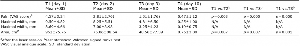 Click to show fullsize